. 2018 Dec 11;11:8995-9006.
doi: 10.2147/OTT.S186247. eCollection 2018.

Resveratrol induces apoptosis in human melanoma cell through negatively regulating Erk/PKM2/Bcl-2 axis

Affiliations
Free PMC article

Resveratrol induces apoptosis in human melanoma cell through negatively regulating Erk/PKM2/Bcl-2 axis

Hailong Zhao et al. Onco Targets Ther. .
Free PMC article

Abstract

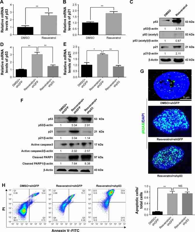
Background: Resveratrol is known as a natural phytoalexin found in grapes and wine, which has significant antitumor activity under in vitro and in vivo conditions. In recent years, great progress has been made in understanding the underlying mechanisms of resveratrol in inducing cellular apoptosis of melanoma cells. Our previous study has shown that the apoptosis regulation of resveratrol in melanoma cells was independent of activation of classical apoptosis-related protein p53.

Materials and methods: MTT assay and 5-bromo-2'-deoxyuridine staining assay were used to analyze cell viability and proliferation. Immunofluorescence analysis of γ-H2AX was employed to clarify DNA damages. Annexin V-propidine iodide/fluorescein isothiocyanate assay was performed to evaluate the cell apoptosis. The mechanisms underlying the activation of M2-type pyruvate kinase (PKM2) by Erk1/2 to stabilize and maintain Bcl-2 signaling was investigated by subcellular fractionation analyses, immunofluorescence analysis, co-immunoprecipitation assay, ubiquitination assay, and glutathione S-transferase pull-down assay.

Results: In the present study, we found that resveratrol dramatically inhibited melanoma cell proliferation and induced cell apoptosis through upregulation of p53 in a concentration-dependent manner. Conversely, p53 downregulation by short hairpin RNA couldn't rescue resveratrol-induced cell proliferation inhibition or apoptosis enlargement. Additionally, we found that resveratrol downregulated antiapoptotic protein Bcl-2 and activated Bax in the protein levels by promoting Bcl-2 degradation and cytochrome c release. Moreover, we discovered that PKM2, had a key role in cell apoptosis triggered by resveratrol through interacting with Bcl-2. Based on these results, we overexpressed PKM2 in melanoma cells and found that this prevented resveratrol-induced apoptosis by stabilizing the protein level of Bcl-2.

Conclusion: Taken together, our results provided a novel mechanism accounting for the apoptosis induction of resveratrol in melanoma cells and suggested that downregulating Erk/PKM2/Bcl-2 axis appears to be a new approach for the prevention or treatment of melanoma.

Keywords: ER stress; antitumor; cytochrome c; ubiquitination.

Conflict of interest statement

Disclosure The authors report no conflicts of interest in this work.

Figures

Figure 1
Resveratrol inhibited the proliferation and induced apoptosis in human melanoma cells. Notes: (A) Cell morphology of MV3 and A375 melanoma cells after treating with the indicated concentration of resveratrol for 48 hours; scale bar as shown. Treatment with DMSO was used as control. (B) MTT assay was performed to examine the effect of indicated concentration of resveratrol on cell viability. All data were shown as the mean ± SD, *P<0.05, **P<0.01, ***P<0.001, and NS P≥0.05. (C) Image and quantification of MV3 cells positive for BrdU staining after treating as in (B); scale bar as shown. (D) Immunofluorescence analysis to identify γH2AX-positive nuclei in MV3 cells treated as in (B). (E) Apoptosis of MV3 cells treated as in (B). Cells were stained with PI and Annexin V–FITC, and the positively stained cells were counted using flow cytometry. Quantification of apoptotic cells is presented on the lower left. (F) Western blot analysis was performed for evaluating the activation of cleaved Caspase3 and expression of cleaved PARP1 in MV3 cells treated as in (B). β-Actin acts as loading control. Densitometry of Western blot in the bottom of each panel is shown as the ratio to β-actin. Abbreviation: BrdU, 5-bromo-2′-deoxyuridine; DMSO, dimethyl sulfoxide; FITC, fluorescein isothiocyanate; NS, not significant; PI, propidine iodide.
Figure 2
Resveratrol-induced apoptosis was independent of the p53-mediated pathway in human melanoma cells. Notes: (A and B) MV3 cells were treated with 200 μM resveratrol or DMSO (as control) for 48 hours. RNA was isolated for performing qPCR using β-actin as reference gene to determine p53 and p21 mRNA levels. All data were shown as the mean±SD, *P<0.05, **P<0.01, and NS P≥0.05. (C) Western blot analysis of the protein levels of p53 and acetylated p53 and p21 in extracts prepared from MV3 cells treated with 200 μM resveratrol or DMSO. Expression of β-actin was used as a loading control. (D and E) mRNA levels of p53 and p21 in MV3 cells treated with DMSO or resveratrol-transduced with indicated shRNA for p53/GFP by RT-qPCR were analyzed. (F) The expression levels of p53, p21, active caspase3, and cleaved PARP1 were determined using Western blot analysis after cells were treated as in (D). (G) Immunofluorescence analysis to identify γH2AX-positive nuclei in MV3 cells treated as in (D). (H) Apoptosis was analyzed in MV3 cells treated as in (D). Quantification of apoptotic cells is presented on the lower right. Abbreviations: DAPI, diamidine phenylindole; DMSO, dimethyl sulfoxide; GFP, green fluorescent protein; NS, not significant; RT-qPCR, real-time quantitative PCR; shGFP, GFP-specific shRNA; shRNA, short hairpin RNA.
Figure 3
Figure 3
Effect of resveratrol involved in degradation of Bcl-2 and release of cytochrome c in human melanoma cells. Notes: (A and B) shGFP- or shp53-transduced MV3 cells were treated with or without resveratrol (200 μM, 48 hours). A qRT-PCR was performed to examine mRNA levels of Bcl-2 and Bax. All data were shown as the mean ± SD, **P<0.01, and NS P≥0.05. (C) The expressions of p53, Bcl-2, and Bax were checked in shp53-transduced MV3 cells treated with or without 200 μM resveratrol for 48 hours. DMSO and shGFP vector were used as control. β-Actin was used as internal reference. (D) Resveratrol-treated and/or shp53-transduced MV3 cells were treated with MG132 for 6 hours before harvesting. The ubiquitinated Bcl-2 proteins were pulled down with anti-HA antibody and immunoblotted with anti-Bcl-2 antibody using streptavidin agarose beads. (E) MV3 cells treated with or without resveratrol were harvested for cytosolic (Cyto) and mitochondrial (Mito) fractionation. Western blot assay was used to show the expression levels of cytochrome c in cytosolic or mitochondrial fraction. VDAC and Tublin were used as loading control. (F) Immunofluorescence analyses were performed using anti-cytochrome c or anti-VDAC antibodies in MV3 cells treated with or without resveratrol. Scale bar as shown. Abbreviations: DAPI, diamidine phenylindole; DMSO, dimethyl sulfoxide; IB, immunoblotting; IP, immunoprecipitation; NS, not significant; qRT-PCR, real-time quantitative PCR; shGFP, GFP-specific shRNA; VDAC, voltage dependent anion channel protein.
Figure 4
PKM2 interacted with Bcl-2 and resisted resveratrol-induced apoptosis in human melanoma cells. Notes: (A) Western blots showing the expression of p-ERK1/2, ERK1/2, PKM2, and Bcl-2 in MV3 cells after treating with 200 μM resveratrol for 48 hours. (B) RNA was extracted from MV3 cells after treating with 200 μM resveratrol for 48 hours and qPCR was performed using indicated primers for analysis of PKM2. All data were shown as the means ± SD, *P<0.05, **P<0.01, and NS P≥0.05. (C) Reciprocal co-IP of PKM2 and Bcl-2 in MV3 melanoma cells followed by Western blot analysis. IgG represents a control antibody used for IPs. Five hundred micrograms of lysates were used for each IP reaction and 50 μg lysates were used as input controls. (D) Immunofluorescence was performed using anti-PKM2 and anti-Bcl-2 antibodies in MV3 cells after treating with 200 μM resveratrol for 48 hours. Scale bar as shown. (E) Direct interaction of recombinant GST-PKM2 protein with Bcl-2 in GST pull-down. GST-tagged PKM2 proteins purified from MV3 cells after treating with resveratrol (200 μM) or TEPP-46 (1 μM) for 48 hours were detected by anti-Bcl-2 antibody (bottom), and protein loading was detected by CBB staining after resolution by 8% SDS-PAGE (top). (F) mRNA levels of PKM2 were analyzed by qPCR in PKM2 overexpressing MV3 cells that were treated with resveratrol as indicated. DMSO and empty vector were used as controls. (G) The expression levels of indicated proteins were analyzed using Western blot assays in PKM2 overexpressing and control cells that were treated with resveratrol as indicated. β-Actin was used as a loading control. (H) Image and quantification of PKM2-overexpressed MV3 cells after treating with resveratrol as well as control cells positive for BrdU staining. Scale bar as shown. (I) Apoptosis was analyzed in MV3 overexpressing with Bcl after treating with resveratrol as indicated. DMSO and empty vector were used as controls. Quantification of apoptotic cells is presented on the lower right. (J) Schema illustrating the mechanism of regulation of resveratrol on PKM2-mediated Bcl-2 stabilization and apoptosis resistance in human melanoma cell. Abbreviations: CBB, coomassie brilliant blue; co-IP, co-immunoprecipitation; DAPI, diamidine phenylindole; DMSO, dimethyl sulfoxide; GST, glutathione S-transferase; PKM2, M2-type pyruvate kinase.

Similar articles

See all similar articles

Cited by 5 articles

References

    1. Rastrelli M, Tropea S, Rossi CR, Alaibac M. Melanoma: epidemiology, risk factors, pathogenesis, diagnosis and classification. In Vivo. 2014;28(6):1005–1011. - PubMed
    1. Hu H, Dong Z, Tan P, et al. Antibiotic drug tigecycline inhibits melanoma progression and metastasis in a p21CIP1/Waf1-dependent manner. Oncotarget. 2016;7(3):3171–3185. - PMC - PubMed
    1. Vijuk G, Coates AS. Survival of patients with visceral metastatic melanoma from an occult primary lesion: a retrospective matched cohort study. Ann Oncol. 1998;9(4):419–422. - PubMed
    1. Neila J, Soyer HP. Key points in dermoscopy for diagnosis of melanomas, including difficult to diagnose melanomas, on the trunk and extremities. J Dermatol. 2011;38(1):3–9. - PubMed
    1. Veierød MB, Weiderpass E, Thörn M, et al. A prospective study of pigmentation, sun exposure, and risk of cutaneous malignant melanoma in women. J Natl Cancer Inst. 2003;95(20):1530–1538. - PubMed
Feedback