. 2014 Dec;76(6):845-61.
doi: 10.1002/ana.24271. Epub 2014 Sep 26.

Impairment of paravascular clearance pathways in the aging brain

Affiliations
Free PMC article

Impairment of paravascular clearance pathways in the aging brain

Benjamin T Kress et al. Ann Neurol. .
Free PMC article

Abstract

Objective: In the brain, protein waste removal is partly performed by paravascular pathways that facilitate convective exchange of water and soluble contents between cerebrospinal fluid (CSF) and interstitial fluid (ISF). Several lines of evidence suggest that bulk flow drainage via the glymphatic system is driven by cerebrovascular pulsation, and is dependent on astroglial water channels that line paravascular CSF pathways. The objective of this study was to evaluate whether the efficiency of CSF-ISF exchange and interstitial solute clearance is impaired in the aging brain.

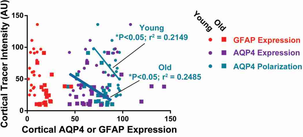
Methods: CSF-ISF exchange was evaluated by in vivo and ex vivo fluorescence microscopy and interstitial solute clearance was evaluated by radiotracer clearance assays in young (2-3 months), middle-aged (10-12 months), and old (18-20 months) wild-type mice. The relationship between age-related changes in the expression of the astrocytic water channel aquaporin-4 (AQP4) and changes in glymphatic pathway function was evaluated by immunofluorescence.

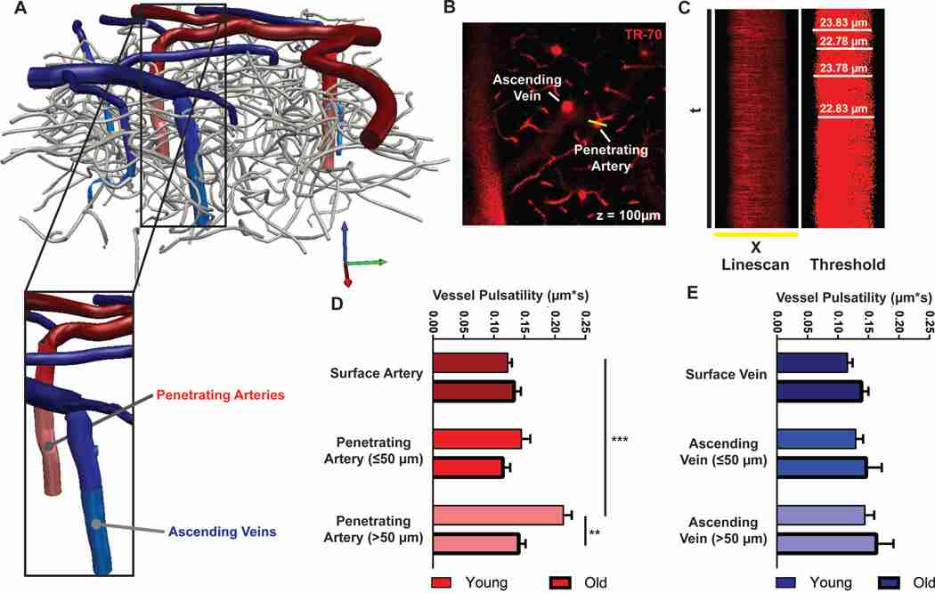
Results: Advancing age was associated with a dramatic decline in the efficiency of exchange between the subarachnoid CSF and the brain parenchyma. Relative to the young, clearance of intraparenchymally injected amyloid-β was impaired by 40% in the old mice. A 27% reduction in the vessel wall pulsatility of intracortical arterioles and widespread loss of perivascular AQP4 polarization along the penetrating arteries accompanied the decline in CSF-ISF exchange.

Interpretation: We propose that impaired glymphatic clearance contributes to cognitive decline among the elderly and may represent a novel therapeutic target for the treatment of neurodegenerative diseases associated with accumulation of misfolded protein aggregates.

Conflict of interest statement

Conflict of Interest: The authors declare that they have no competing conflict of interest

Figures

Figure 1. Age-dependent decline in paravascular glymphatic CSF recirculation and interstitial solute efflux
(A) Small molecular weight (Texas Red-conjugated dextran, 3 kD; Dex-3) and large molecular weight tracer (ovalbumin-conjugated ALEXA-647, 45 kD; OA-45) were injected intracisternally into young (2–3 months), middle age (10–12 months) and old (18 months) mice. (B) 30 min after injection, animals were perfusion fixed and whole-slice fluorescence was evaluated. CSF tracer penetration was significantly reduced in the old brain compared to the young brain, while values in the middle age brains were intermediate (*P<0.05, ***P<0.001; 1-way ANOVA; n = 5–8 per group). (C) Representative images show that compared to young brains, CSF tracer penetration into middle age and old brains is markedly slowed. (D) Radio-labeled 125I-amyloid β1–40 and 14C-Inulin were co-injected into the caudate nucleus of young, middle aged and old mice. 60 min after injection, solute clearance was evaluated by gamma counting and liquid scintillation counting. (E) Compared to clearance in the young brain, 125I-amyloid β1–40 and 14C-Inulin clearance were significantly impaired in the old month brain (***P<0.001, **P<0.01, *P<0.05, 1-way ANOVA; n = 6–11 per group). 125I-amyloid β1–40 and 14C-Inulin clearance in the middle age brain were intermediate between that of the young and old brain.
Figure 2. In vivo 2-photon imaging reveals suppressed paravascular glymphatic CSF recirculation in aging mice
(A) Paravascular CSF tracer penetration into the mouse cortex was evaluated by in vivo 2-photon microscopy after intracisternal injection of FITC-conjugated dextran (40 kD, Dex-40). (B) Quantification of CSF tracer penetration 100 µm below the cortical surface shows impaired paravascular penetration in middle age compared to young cortex (*P<0.05, 2-way repeated measures ANOVA; n = 4 animals per group). (C-D) Serial in vivo 2-photon imaging at the cortical surface and 100 µm below the cortical surface after intracisternal Dex-40 injection. Cerebral vasculature is visualized by intra-arterial injection of Texas Red-conjugated dextran (70 kD, Dex-70, blue); surface arteries (arrows) and veins (arrowheads) are defined morphologically. Compared to the young brain (C), CSF tracer movement along the surface and penetrating arteries and into the surrounding interstitium is markedly slowed in the middle age brain (D).
Figure 3. Vascular pulsatility is suppressed in the penetrating arteries of the aging brain
Pulsatility of the vascular wall was evaluated by in vivo 2-photon microscopy through thin-skull cranial window after intra-arterial injection of Texas Red-conjugated dextran (MW 70kD, dex-70). (A) 3D reconstruction of the cerebrovascular tree visible through cranial window reveals penetrating arteries (red), ascending veins (blue) and intervening capillary bed (gray). (B) Between z = 0 and 150 µm below the cortical surface, high-frequency orthogonal linescans were generated across surface and penetrating arteries and surface and ascending veins. (C) Representative raw x-t scans were thresholded to improve edge detection, and the luminal diameter was measured over time. (D-E) Vascular pulsatility was measured from penetrating arteries and ascending veins. In the young brain, arterial pulsatility was significantly greater than venous pulsatility (*P<0.05, 2-way ANOVA; n = 8–20 vessels from 4 animals per group). In the old brain, arterial pulsatility in the penetrating arteries was significantly reduced compared to the young brain (*P<0.05, Young vs. Old). No age-related differences in venous pulsatility were observed.
Figure 4. Interstitial volume does not change as a function of aging
(A) Extracellular volume fraction (α) and tortuosity (λ) were evaluated by the in vivo TMA micro-iontophoresis method. TMA is introduced by an iontophoresis electrode and detected by a second recording electrode. Changes in the extracellular volume fraction or the tortuosity of the extracellular space are reflected in the kinetics of the measured TMA concentrations (described in detail in(18)). (B) In the cortex of either awake or anesthetized mice, extracellular volume fraction did not significantly differ between young and old animals. Both young and old brains exhibited a significant and comparable enlargement of the extracellular space in the anesthetized state (***P<0.001, Awake vs. Anesthetized; 2-Way ANOVA; n = 9–20 per group). (C) Extracellular tortuosity did not differ significantly between young and old animals.
Figure 5. Perivascular AQP4 polarization surrounding penetrating arteries is lost in the aging brain
Changes in AQP4 localization, GFAP expression and paravascular CSF recirculation were evaluated by immunofluorescence double-labeling. (A-B) Slices from brains fixed 30 min after intracisternal injection of fluorescent CSF tracer ovalbumin-conjugated ALEXA-647 (MW 45kD, OA-45) were imaged by confocal microscopy and show that compared to the young cortex (A), AQP4 localization becomes more disperse in the old brain while GFAP expression increases and CSF tracer penetration is slowed (B). AQP4 and GFAP immunofluorescence were evaluated in linear regions of interest (dashed lines) extending outward from penetrating cerebral arterioles (arrowheads, C, F) or outward from cerebral capillaries (arrows, D, G). (C-D, F-G) Compared to the young brain, and GFAP expression were increased surrounding blood vessels in the aging brain (***P<0.001, young vs. old; repeated measures 2-way ANOVA; n = 11–18 vessels from 4 animals per group). Changes in AQP4 expression were greater surrounding penetrating arterioles (C) than cortical capillaries (B), with marked upregulation of AQP4 in tissue surrounding penetrating vessels. Measurement of perivascular AQP4 (E) and GFAP (H) expression showed that AQP4 is downregulated in perivascular domains surrounding penetrating arterioles, but not around capillaries (*P< 0.05; un-paired t-test, n = 11–18 vessels from 4 animals per group) while perivascular GFAP expression was similarly upregulated surrounding both vessel types (***P<0.001; un-paired t-test).
Figure 6. Impairment of perivascular AQP4 polarization is greatest in the lateral and ventral cortex, hippocampus and striatum of the aging brain
AQP4 expression and polarization and GFAP expression were evaluated by immunofluorescence in fixed brain slices from young (2–3 month) and old (18 month) brains. Expression and polarization were evaluated within different regions of anterior (A-C) and posterior (D-F) brain slices. Regional heat maps depict mean change in AQP4 expression (AQP4 immunofluorescence), AQP4 polarization (% area), and GFAP expression (% area) between young and old brains. (A, D) Within both the anterior and posterior regions, global AQP4 immunofluorescence did not differ between the young and old brains. (B, E) Perivascular AQP4 polarization was significantly reduced in the aged brain, with most pronounced effects in the lateral and ventral cortex, striatum and hippocampus (*P<0.05, **P<0.01, ***<0.001, Young vs. Old; 2-way ANOVA; n = 4 per group). (C, F) GFAP expression was significantly increased in the old compared to the young brain, with the greatest effect evident within the lateral and ventral cortex, hippocampus and striatum (*P<0.05, **P<0.01, ***<0.001, Young vs. Old; 2-way ANOVA; n = 4 per group).
Figure 7. Ventral and lateral cortex exhibit the most severe suppression of glymphatic pathway activity in the aged brain
Age-related impairment of paravascular CSF recirculation was evaluated 30min after intracisternal injection of small molecular weight CSF tracer Texas Red-conjugated dextran (MW 3kD, Dex-3) and large molecular weight ovalbumin-conjugated ALEXA 647 (MW 45kD, OA-45). Representative saggital and coronal slices from young (A) and old (B) brains show marked reduction in CSF tracer penetration throughout the aged compared to the young brain. CSF tracer penetration within defined brain regions was evaluated and heat maps from anterior (C) and posterior (D) slices depict the change in mean OA-45 CSF tracer penetration observed in each region between old and young brains. Quantification of OA-45 penetration in different regions showed that paravascular CSF recirculation was significantly impaired in the lateral and ventral cortex of both the anterior and posterior slices (*P<0.05, **P<0.01, ***P<0.001, Young vs. Old; 2-way ANOVA; n = 4 per group).
Figure 8. Loss of cortical perivascular AQP4 polarization is associated with impaired paravascular CSF recirculation
(A) The association between AQP4 expression, AQP4 polarization or GFAP expression and paravascular CSF tracer was evaluated in the cortex. Linear regression analysis shows that GFAP and AQP4 expression levels were not associated with differences in paravascular CSF tracer penetration in the young or old cortex. Within both the young and old cortex, reduction in perivascular AQP4 polarization were significantly associated with impairment of paravascular CSF recirculation (Young brain: *P<0.05, r2 = 0.2149; Old brain: *P<0.05, r2 = 0.2485).
Figure 9. Increased pial surface AQP4 expression is associated with impaired trans-pial CSF penetration
The association between changes in subpial AQP4 and GFAP expression and trans-pial CSF tracer penetration were evaluated in the lateral cerebral cortex of young (A) and old (B) mice. (C) AQP4 and GFAP expression and CSF tracer intensity were evaluated in linear regions of interest (white dashed lines) extending inward from the pial surface. (D) Compared to the young brain, AQP4 expression in the aging brain was significantly greater near the pial surface (**P<0.01, Young vs. Old; 2-way repeated measures ANOVA, n = 6–8 per group) while GFAP immunoreactivity was not significantly altered. Increased sub-pial AQP4 expression in the aging brain was associated with a significant reduction in trans-pial CSF tracer penetration (**P<0.01, Young vs. Old; 2-way repeated measures ANOVA, n = 6–8 per group).

Similar articles

See all similar articles

Cited by 247 articles

See all "Cited by" articles

Publication types

Feedback