Cerebrospinal fluid is drained primarily via the spinal canal and olfactory route in young and aged spontaneously hypertensive rats
- PMID: 24932405
- PMCID: PMC4057524
- DOI: 10.1186/2045-8118-11-12
Cerebrospinal fluid is drained primarily via the spinal canal and olfactory route in young and aged spontaneously hypertensive rats
Abstract
Background: Many aspects of CSF dynamics are poorly understood due to the difficulties involved in quantification and visualization. In particular, there is debate surrounding the route of CSF drainage. Our aim was to quantify CSF flow, volume, and drainage route dynamics in vivo in young and aged spontaneously hypertensive rats (SHR) using a novel contrast-enhanced computed tomography (CT) method.
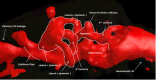
Methods: ICP was recorded in young (2-5 months) and aged (16 months) SHR. Contrast was administered into the lateral ventricles bilaterally and sequential CT imaging was used to visualize the entire intracranial CSF system and CSF drainage routes. A customized contrast decay software module was used to quantify CSF flow at multiple locations.
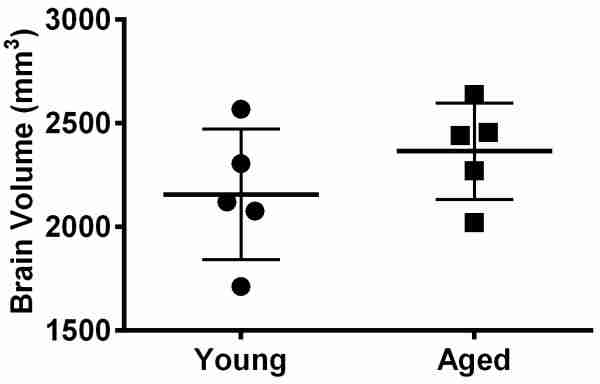
Results: ICP was significantly higher in aged rats than in young rats (11.52 ± 2.36 mmHg, versus 7.04 ± 2.89 mmHg, p = 0.03). Contrast was observed throughout the entire intracranial CSF system and was seen to enter the spinal canal and cross the cribriform plate into the olfactory mucosa within 9.1 ± 6.1 and 22.2 ± 7.1 minutes, respectively. No contrast was observed adjacent to the sagittal sinus. There were no significant differences between young and aged rats in either contrast distribution times or CSF flow rates. Mean flow rates (combined young and aged) were 3.0 ± 1.5 μL/min at the cerebral aqueduct; 3.5 ± 1.4 μL/min at the 3rd ventricle; and 2.8 ± 0.9 μL/min at the 4th ventricle. Intracranial CSF volumes (and as percentage total brain volume) were 204 ± 97 μL (8.8 ± 4.3%) in the young and 275 ± 35 μL (10.8 ± 1.9%) in the aged animals (NS).
Conclusions: We have demonstrated a contrast-enhanced CT technique for measuring and visualising CSF dynamics in vivo. These results indicate substantial drainage of CSF via spinal and olfactory routes, but there was little evidence of drainage via sagittal sinus arachnoid granulations in either young or aged animals. The data suggests that spinal and olfactory routes are the primary routes of CSF drainage and that sagittal sinus arachnoid granulations play a minor role, even in aged rats with higher ICP.
Keywords: Age; CSF; Cerebrospinal fluid dynamics; Computed tomography; Contrast; Intracranial pressure (ICP); SHR; Spontaneously hypertensive rat.
Figures
Similar articles
-
Cerebrospinal fluid circulation and associated intracranial dynamics. A radiologic investigation using MR imaging and radionuclide cisternography.Acta Radiol Suppl. 1993;386:1-23. Acta Radiol Suppl. 1993. PMID: 8517189
-
Cerebrospinal fluid outflow resistance in sheep: impact of blocking cerebrospinal fluid transport through the cribriform plate.Neuropathol Appl Neurobiol. 2002 Feb;28(1):67-74. doi: 10.1046/j.1365-2990.2002.00373.x. Neuropathol Appl Neurobiol. 2002. PMID: 11849565
-
New concept of cerebrospinal fluid dynamics in cerebral venous sinus thrombosis.Med Hypotheses. 2008;70(1):143-7. doi: 10.1016/j.mehy.2007.03.036. Epub 2007 Jun 13. Med Hypotheses. 2008. PMID: 17570605
-
Lymphatic drainage of cerebrospinal fluid in mammals - are arachnoid granulations the main route of cerebrospinal fluid outflow?Biologia (Bratisl). 2018;73(6):563-568. doi: 10.2478/s11756-018-0074-x. Epub 2018 Jun 27. Biologia (Bratisl). 2018. PMID: 30147112 Free PMC article. Review.
-
Anatomy and physiology of cerebrospinal fluid.Eur Ann Otorhinolaryngol Head Neck Dis. 2011 Dec;128(6):309-16. doi: 10.1016/j.anorl.2011.03.002. Epub 2011 Nov 18. Eur Ann Otorhinolaryngol Head Neck Dis. 2011. PMID: 22100360 Review.
Cited by 30 articles
-
Location matters: highly divergent protein levels in samples from different CNS compartments in a clinical trial of rituximab for progressive MS.Fluids Barriers CNS. 2020 Jul 29;17(1):49. doi: 10.1186/s12987-020-00205-4. Fluids Barriers CNS. 2020. PMID: 32727487 Free PMC article.
-
Magnetic Resonance Imaging and Modeling of the Glymphatic System.Diagnostics (Basel). 2020 May 27;10(6):344. doi: 10.3390/diagnostics10060344. Diagnostics (Basel). 2020. PMID: 32471025 Free PMC article. Review.
-
Repetitive Mild Traumatic Brain Injury Alters Glymphatic Clearance Rates in Limbic Structures of Adolescent Female Rats.Sci Rep. 2020 Apr 10;10(1):6254. doi: 10.1038/s41598-020-63022-7. Sci Rep. 2020. PMID: 32277097 Free PMC article.
-
A Study of Antidepressant Effect and Mechanism on Intranasal Delivery of BDNF-HA2TAT/AAV to Rats with Post-Stroke Depression.Neuropsychiatr Dis Treat. 2020 Mar 4;16:637-649. doi: 10.2147/NDT.S227598. eCollection 2020. Neuropsychiatr Dis Treat. 2020. PMID: 32184603 Free PMC article.
-
Vital Functions Contribute to the Spread of Extracellular Fluids in the Brain: Comparison Between Life and Death.Front Aging Neurosci. 2020 Feb 11;12:15. doi: 10.3389/fnagi.2020.00015. eCollection 2020. Front Aging Neurosci. 2020. PMID: 32116648 Free PMC article.
References
-
- Wagshul ME, McAllister JP, Rashid S, Li J, Egnor MR, Walker ML, Yu M, Smith SD, Zhang G, Chen JJ, Benveniste H. Ventricular dilation and elevated aqueductal pulsations in a new experimental model of communicating hydrocephalus. Exp Neurol. 2009;218:33–40. doi: 10.1016/j.expneurol.2009.03.034. - DOI - PubMed
LinkOut - more resources
-
Full Text Sources
-
Other Literature Sources
-
Medical
-
Research Materials
-
Miscellaneous
