Basic vascular neuroanatomy of the brain and spine: what the general interventional radiologist needs to know
- PMID: 24436544
- PMCID: PMC3773035
- DOI: 10.1055/s-0033-1353475
Basic vascular neuroanatomy of the brain and spine: what the general interventional radiologist needs to know
Abstract
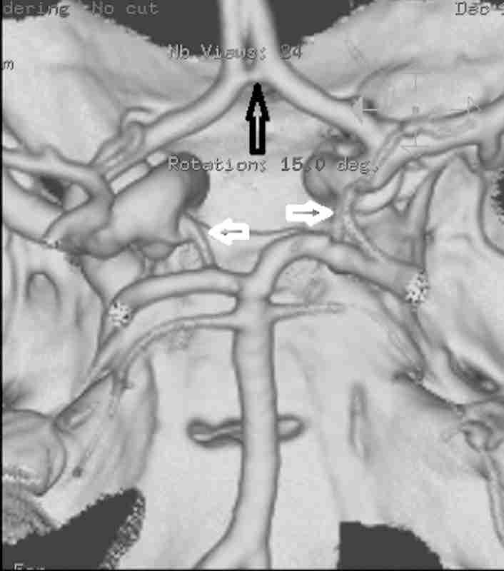
This article is intended to provide a review of clinically relevant neurovascular anatomy. A solid understanding of the vascular anatomy of the brain and spine are essential for the safe and effective performance of neurointerventional radiology. Key concepts to master include collateral pathways and anastomoses between the external and internal carotid circulation, the Circle of Willis as a route to otherwise inaccessible intracranial vascular distributions, and the origin of spinal arterial blood supply. These concepts will be highlighted using clinical angiographic examples with discussion of relevant embryology and pathology as needed.
Keywords: angiography; interventional radiology; neuroanatomy; neurointerventional radiology.
Figures
Similar articles
-
Dangerous Extracranial-Intracranial Anastomoses: What the Interventionalist Must Know.Semin Intervent Radiol. 2020 Jun;37(2):140-149. doi: 10.1055/s-0040-1709155. Epub 2020 May 14. Semin Intervent Radiol. 2020. PMID: 32419726 Review.
-
Common Cervical and Cerebral Vascular Variants.Interv Cardiol Clin. 2014 Jan;3(1):123-134. doi: 10.1016/j.iccl.2013.09.002. Epub 2013 Nov 26. Interv Cardiol Clin. 2014. PMID: 28582148 Review.
-
Intracranial collateral anastomoses: relevance to endovascular procedures.Neurosurg Clin N Am. 2009 Jul;20(3):279-96. doi: 10.1016/j.nec.2009.04.013. Neurosurg Clin N Am. 2009. PMID: 19778700 Review.
-
A Functional Perspective on the Embryology and Anatomy of the Cerebral Blood Supply.J Stroke. 2015 May;17(2):144-58. doi: 10.5853/jos.2015.17.2.144. Epub 2015 May 29. J Stroke. 2015. PMID: 26060802 Free PMC article. Review.
-
Presentation of variations in the anterior part of the circle of Willis as a result of MRI-angiography method.Med Arh. 2004;58(6):327-30. Med Arh. 2004. PMID: 15648225
Cited by 9 articles
-
Bioprinting of freestanding vascular grafts and the regulatory considerations for additively manufactured vascular prostheses.Transl Res. 2019 Sep;211:123-138. doi: 10.1016/j.trsl.2019.05.005. Epub 2019 Jun 3. Transl Res. 2019. PMID: 31201778 Review.
-
Posthemorrhagic hydrocephalus development after germinal matrix hemorrhage: Established mechanisms and proposed pathways.J Neurosci Res. 2020 Jan;98(1):105-120. doi: 10.1002/jnr.24394. Epub 2019 Feb 21. J Neurosci Res. 2020. PMID: 30793349 Free PMC article. Review.
-
Magnetic resonance angiography determined variations in the circle of Willis: Analysis of a large series from a single center.Ci Ji Yi Xue Za Zhi. 2019 Jan-Mar;31(1):52-59. doi: 10.4103/tcmj.tcmj_167_17. Ci Ji Yi Xue Za Zhi. 2019. PMID: 30692833 Free PMC article.
-
Toward dynamic lumbar puncture guidance using needle-based single-element ultrasound imaging.J Med Imaging (Bellingham). 2018 Apr;5(2):021224. doi: 10.1117/1.JMI.5.2.021224. Epub 2018 Apr 2. J Med Imaging (Bellingham). 2018. PMID: 29651451 Free PMC article.
-
The Paravascular Pathway for Brain Waste Clearance: Current Understanding, Significance and Controversy.Front Neuroanat. 2017 Nov 7;11:101. doi: 10.3389/fnana.2017.00101. eCollection 2017. Front Neuroanat. 2017. PMID: 29163074 Free PMC article. Review.
