. 2013 Nov 13;33(46):18190-9.
doi: 10.1523/JNEUROSCI.1592-13.2013.

Cerebral arterial pulsation drives paravascular CSF-interstitial fluid exchange in the murine brain

Affiliations
Free PMC article

Cerebral arterial pulsation drives paravascular CSF-interstitial fluid exchange in the murine brain

Jeffrey J Iliff et al. J Neurosci. .
Free PMC article

Abstract

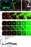
CSF from the subarachnoid space moves rapidly into the brain along paravascular routes surrounding penetrating cerebral arteries, exchanging with brain interstitial fluid (ISF) and facilitating the clearance of interstitial solutes, such as amyloid β, in a pathway that we have termed the "glymphatic" system. Prior reports have suggested that paravascular bulk flow of CSF or ISF may be driven by arterial pulsation. However, cerebral arterial pulsation could not be directly assessed. In the present study, we use in vivo two-photon microscopy in mice to visualize vascular wall pulsatility in penetrating intracortical arteries. We observed that unilateral ligation of the internal carotid artery significantly reduced arterial pulsatility by ~50%, while systemic administration of the adrenergic agonist dobutamine increased pulsatility of penetrating arteries by ~60%. When paravascular CSF-ISF exchange was evaluated in real time using in vivo two-photon and ex vivo fluorescence imaging, we observed that internal carotid artery ligation slowed the rate of paravascular CSF-ISF exchange, while dobutamine increased the rate of paravascular CSF-ISF exchange. These findings demonstrate that cerebral arterial pulsatility is a key driver of paravascular CSF influx into and through the brain parenchyma, and suggest that changes in arterial pulsatility may contribute to accumulation and deposition of toxic solutes, including amyloid β, in the aging brain.

Figures

Figure 1.
Figure 1.
CSF enters the brain along para-arterial pathways. A, Fluorescent tracer (OA-647; MW, 45 kDa) injected intracisternally into the subarachnoid CSF enters the brain parenchyma along paravascular pathways. B, Ex vivo confocal imaging in NG2-DsRed mice showed that 15 min after injection, CSF tracer enters the brain along DsRed-positive penetrating arteries (hollow arrowheads), but not along DsRed-negative ascending veins (filled arrowheads). C, In vivo two-photon imaging in NG2-DsRed mice after fluorescence angiography (intravenous FITC-conjugated dextran-2000; MW, 2000 kDa) shows that cortical arteries and veins can be readily distinguished in these mice. D–G, Time-lapse in vivo two-photon imaging of florescent CSF tracer (FITC-d40; MW, 40 kDa) influx into the cortex after intracisternal injection. Cerebral vasculature is imaged by intra-arterial injection of Texas Red-conjugated dextran-70 (TR-d70; MW, 70 kDa). D, At the cortical surface, CSF tracer moves via para-arterial spaces. E, F, 100 μm below the cortical surface, CSF tracer enters the cortex along penetrating arteries, then exchanges with the surrounding interstitium. G, After para-arterial influx, CSF tracer is also evident along ascending veins. H, Measurement of ICP during intracisternal CSF tracer infusion (1 μl/min for 10 min) resulted in mild (∼2.5 mmHg) elevation of ICP that resolved rapidly upon cessation of infusion.
Figure 2.
Measurement of vascular pulsatility by in vivo two-photon imaging. A, The cerebral vasculature was visualized by in vivo two-photon fluorescence angiography after intravenous injection of FITC-conjugated detran-2000 (FITC-d2000; MW, 2000 kDa). B, C, Cortical surface arteries and veins (B), and penetrating arteries and ascending veins (C) were selected and X–T line scans (orange lines) were generated orthogonal to the vessel axis. D, E, Vessel diameter was measured and plotted as a function of time. Vascular pulsatility was defined as the absolute value of the integral of the vascular diameter approximately a running average over a 3000 ms epoch. F, Vascular pulsatility was measured in cortical surface arteries (SA), penetrating arteries (PA), ascending veins (AV), and surface veins (SV). Pulsatility was greatest in penetrating arteries and veins compared with surface vessels.
Figure 3.
Reduced cortical arterial pulsatility after internal carotid artery ligation. A, Unilateral internal carotid artery ligation did not appreciably alter systemic blood pressure. B, Immediately following carotid artery ligation, CBF measured by LDF was reduced ∼25%, recovering to baseline values within 30 min. C, When imaged via an in vivo cranial window preparation, cerebral vessel wall pulsatility was significantly reduced in penetrating cortical arteries, but remained unchanged in surface arteries, ascending veins, and surface veins (**p < 0.01 sham vs ligation; 2-way ANOVA; n = 16–45 vessels per group). D, E, Unilateral internal carotid artery ligation did not significantly alter the diameter of cortical surface arteries, penetrating arteries, ascending veins, surface veins, or heart rate. F, When imaged by a thin-skull preparation that leaves the skull intact, internal carotid artery ligation significantly reduced cerebral arterial wall pulsatility in surface arteries, penetrating arteries, and ascending veins (*p < 0.05 sham vs ligation; 2-way ANOVA; n = 12–15 vessels per group). G, H, Carotid artery ligation did not alter cortical vascular diameter or heart rate when imaging was conducted through a thin-skull preparation.
Figure 4.
Internal carotid artery ligation reduces paravascular CSF influx. A, B, Paravascular influx of CSF tracer (FITC-d40; MW, 40 kDa) was first evaluated by in vivo two-photon microscopy at the cortical surface, 100 and 160 μm below the cortical surface in sham (A) animals and those undergoing unilateral carotid artery ligation (B). C, Quantification of paravascular CSF tracer 100 μm below the surface demonstrated that carotid artery ligation significantly slowed CSF tracer influx (*p < 0.05, **p < 0.01; 2-way repeated-measures ANOVA; n = 6 animals per group). D, A similar reduction in CSF tracer penetration into the surrounding parenchyma was observed 100 μm below the cortical surface (*p < 0.05; 2-way repeated-measures ANOVA; n = 6 animals per group). E, F, CSF tracer (OA-647; MW, 45 kDa) influx into deeper brain tissue was evaluated by ex vivo fluorescence imaging 30 min after intracisternal tracer injection. G, Quantification of fluorescence intensity in anterior [+0.5 mm − (−1.0) mm relative to bregma] brain regions showed that compared with sham-treated animals, CSF tracer influx was reduced ipsilateral to carotid artery ligation, but not contralateral (*p < 0.05, sham vs ipsilateral side; 2-way ANOVA; n = 4–6 animals per group). H, A greater reduction was evident in posterior [−1.0 mm − (−2.5) mm relative to bregma] brain regions (*p < 0.05, **p < 0.01, ***p < 0.001, sham vs ipsilateral side; 2-way ANOVA; n = 4–6 animals per group). Ctx, Cerebral cortex; WM, subcortical white matter; HC, hippocampus; SCtx, subcortical structures, including striatum, thalamus, and hypothalamus.
Figure 5.
Increased arterial pulsation after systemic dobutamine administration. The effect of systemic dobutamine (40 μg/kg, i.v.) on cortical vessel pulsatility was evaluated by in vivo two-photon angiography 30 min after administration. A, Raw arterial blood pressure trace following systemic dobutamine administration shows an acute effect of the drug that peaks within 10 min of injection and largely resolves over the first 30 min postinjection (the time when intracisternal tracer injections took place). B, Systolic (BPS), diastolic (BPD), and mean (BPMean) arterial blood pressure were significantly increased immediately after systemic dobutamine injection (*p < 0.05 vs baseline values, repeated-measures 1-way ANOVA; n = 8 per group). However, this response resolved within the first 30 min postinjection. C, Heart rate was not altered during the peak blood pressure response to dobutamine. However, 30 min after dobutamine injection, heart rate was significantly elevated compared with baseline values (*p < 0.05, repeated-measures 1-way ANOVA; n = 8 per group). D, 30 min after systemic dobutamine injection, vessel pulsatility was significantly increased only within penetrating arteries but not within other vessel types (*p < 0.05, 2-way ANOVA; n = 10–42 vessels per group). E, Dobutamine did not significantly alter the diameter of surface arteries (SA), penetrating arteries (PA), ascending veins (AV), or surface veins (SV).
Figure 6.
Systemic dobutamine administration increases paravascular CSF tracer influx. The effect of systemic dobutamine administration upon paravascular influx of subarachnoid CSF tracer was evaluated by ex vivo fluorescence imaging 30 min after intracisternal tracer injection. A, B, Compared with sham animals (A), animals treated with dobutamine 30 min before the start of tracer infusion exhibited a marked increase in the influx of CSF tracer into brain tissue (B). C, D, Quantification of CSF tracer influx into anterior (C) and posterior (D) brain regions demonstrated that systemic dobutamine administration significantly increased CSF tracer influx in the parenchyma (*p < 0.05, 2-way ANOVA; n = 4–6 animals per group). Ctx, Cortex; WM, subcortical white matter; HC, hippocampus; SCtx, subcortical structures.

Similar articles

See all similar articles

Cited by 218 articles

See all "Cited by" articles

Publication types

Feedback