Neuroimaging of structural pathology and connectomics in traumatic brain injury: Toward personalized outcome prediction
- PMID: 24179732
- PMCID: PMC3757727
- DOI: 10.1016/j.nicl.2012.08.002
Neuroimaging of structural pathology and connectomics in traumatic brain injury: Toward personalized outcome prediction
Abstract
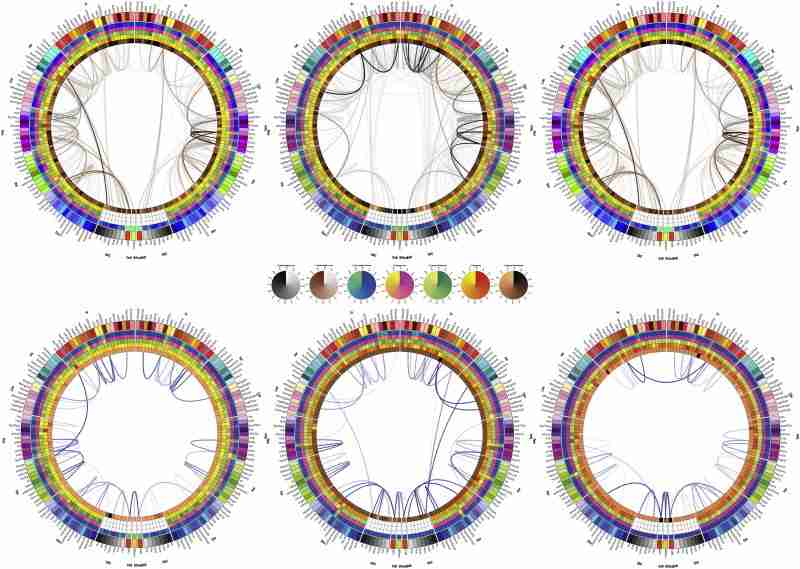
Recent contributions to the body of knowledge on traumatic brain injury (TBI) favor the view that multimodal neuroimaging using structural and functional magnetic resonance imaging (MRI and fMRI, respectively) as well as diffusion tensor imaging (DTI) has excellent potential to identify novel biomarkers and predictors of TBI outcome. This is particularly the case when such methods are appropriately combined with volumetric/morphometric analysis of brain structures and with the exploration of TBI-related changes in brain network properties at the level of the connectome. In this context, our present review summarizes recent developments on the roles of these two techniques in the search for novel structural neuroimaging biomarkers that have TBI outcome prognostication value. The themes being explored cover notable trends in this area of research, including (1) the role of advanced MRI processing methods in the analysis of structural pathology, (2) the use of brain connectomics and network analysis to identify outcome biomarkers, and (3) the application of multivariate statistics to predict outcome using neuroimaging metrics. The goal of the review is to draw the community's attention to these recent advances on TBI outcome prediction methods and to encourage the development of new methodologies whereby structural neuroimaging can be used to identify biomarkers of TBI outcome.
Keywords: 3D, three-dimensional; AAL, Automatic Anatomical Labeling; ADC, apparent diffusion coefficient; ANTS, Advanced Normalization ToolS; BOLD, blood oxygen level dependent; CC, corpus callosum; CT, computed tomography; DAI, diffuse axonal injury; DSI, diffusion spectrum imaging; DTI, diffusion tensor imaging; DWI, diffusion weighted imaging; Diffusion tensor; FA, fractional anisotropy; FLAIR, Fluid Attenuated Inversion Recovery; FSE, Functional Status Examination; GCS, Glasgow Coma Score; GM, gray matter; GOS, Glasgow Outcome Score; GRE, Gradient Recalled Echo; HARDI, high-angular-resolution diffusion imaging; IBA, Individual Brain Atlas; LDA, linear discriminant analysis; MRI, magnetic resonance imaging; MRI/fMRI; NINDS, National Institute of Neurological Disorders and Stroke; Neuroimaging; Outcome measures; PCA, principal component analysis; PROMO, PROspective MOtion Correction; SPM, Statistical Parametric Mapping; SWI, Susceptibility Weighted Imaging; TBI, traumatic brain injury; TBSS, tract-based spatial statistics; Trauma; WM, white matter; fMRI, functional magnetic resonance imaging.
Figures
Similar articles
-
Neuropathology of Mild Traumatic Brain Injury: Correlation to Neurocognitive and Neurobehavioral Findings.In: Kobeissy FH, editor. Brain Neurotrauma: Molecular, Neuropsychological, and Rehabilitation Aspects. Boca Raton (FL): CRC Press/Taylor & Francis; 2015. Chapter 31. Brain Neurotrauma: Molecular, Neuropsychological, and Rehabilitation Aspects. 2015. PMID: 26269912 Free Books & Documents. Review.
-
Traumatic axonal injury: the prognostic value of lesion load in corpus callosum, brain stem, and thalamus in different magnetic resonance imaging sequences.J Neurotrauma. 2014 Sep 1;31(17):1486-96. doi: 10.1089/neu.2013.3258. Epub 2014 Jul 1. J Neurotrauma. 2014. PMID: 24773587
-
Magnetic Resonance Imaging Application in the Area of Mild and Acute Traumatic Brain Injury: Implications for Diagnostic Markers?In: Kobeissy FH, editor. Brain Neurotrauma: Molecular, Neuropsychological, and Rehabilitation Aspects. Boca Raton (FL): CRC Press/Taylor & Francis; 2015. Chapter 24. Brain Neurotrauma: Molecular, Neuropsychological, and Rehabilitation Aspects. 2015. PMID: 26269902 Free Books & Documents. Review.
-
Exploring Serum Biomarkers for Mild Traumatic Brain Injury.In: Kobeissy FH, editor. Brain Neurotrauma: Molecular, Neuropsychological, and Rehabilitation Aspects. Boca Raton (FL): CRC Press/Taylor & Francis; 2015. Chapter 22. Brain Neurotrauma: Molecular, Neuropsychological, and Rehabilitation Aspects. 2015. PMID: 26269900 Free Books & Documents. Review.
-
High angular resolution diffusion-weighted imaging in mild traumatic brain injury.Neuroimage Clin. 2016 Nov 17;13:174-180. doi: 10.1016/j.nicl.2016.11.016. eCollection 2017. Neuroimage Clin. 2016. PMID: 27981032 Free PMC article.
Cited by 41 articles
-
Neuroimaging and Psychometric Assessment of Mild Cognitive Impairment After Traumatic Brain Injury.Front Psychol. 2020 Jul 7;11:1423. doi: 10.3389/fpsyg.2020.01423. eCollection 2020. Front Psychol. 2020. PMID: 32733322 Free PMC article. Review.
-
Investigating Brain Network Changes and Their Association With Cognitive Recovery After Traumatic Brain Injury: A Longitudinal Analysis.Front Neurol. 2020 Jun 9;11:369. doi: 10.3389/fneur.2020.00369. eCollection 2020. Front Neurol. 2020. PMID: 32581989 Free PMC article.
-
Imaging biomarkers of posttraumatic epileptogenesis.Epilepsia. 2019 Nov;60(11):2151-2162. doi: 10.1111/epi.16357. Epub 2019 Oct 8. Epilepsia. 2019. PMID: 31595501 Review.
-
Neuroimaging of traumatic brain injury in military personnel: An overview.J Clin Neurosci. 2019 Dec;70:1-10. doi: 10.1016/j.jocn.2019.07.001. Epub 2019 Jul 19. J Clin Neurosci. 2019. PMID: 31331746 Review.
-
Longitudinal increases in structural connectome segregation and functional connectome integration are associated with better recovery after mild TBI.Hum Brain Mapp. 2019 Oct 15;40(15):4441-4456. doi: 10.1002/hbm.24713. Epub 2019 Jul 11. Hum Brain Mapp. 2019. PMID: 31294921 Free PMC article.
References
-
- Andrews P.J., Sleeman D.H., Statham P.F., McQuatt A., Corruble V., Jones P.A., Howells T.P., Macmillan C.S. Predicting recovery in patients suffering from traumatic brain injury by using admission variables and physiological data: a comparison between decision tree analysis and logistic regression. Journal of Neurosurgery. 2002;97:326–336. - PubMed
-
- Asikainen I., Kaste M., Sarna S. Early and late posttraumatic seizures in traumatic brain injury rehabilitation patients: brain injury factors causing late seizures and influence of seizures on long-term outcome. Epilepsia. 1999;40:584–589. - PubMed
-
- Bazarian J.J., Zhong J., Blyth B., Zhu T., Kavcic V., Peterson D. Diffusion tensor imaging detects clinically important axonal damage after mild traumatic brain injury: a pilot study. Journal of Neurotrauma. 2007;24:1447–1459. - PubMed
