Paravascular microcirculation facilitates rapid lipid transport and astrocyte signaling in the brain
- PMID: 24002448
- PMCID: PMC3761080
- DOI: 10.1038/srep02582
Paravascular microcirculation facilitates rapid lipid transport and astrocyte signaling in the brain
Abstract
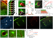
In the brain, a paravascular space exists between vascular cells and astroglial end-foot processes, creating a continuous sheath surrounding blood vessels. Using in vivo two-photon imaging we demonstrate that the paravascular circulation facilitates selective transport of small lipophilic molecules, rapid interstitial fluid movement and widespread glial calcium signaling. Depressurizing the paravascular system leads to unselective lipid diffusion, intracellular lipid accumulation and pathological signaling in astrocytes. As the central nervous system is devoid of lymphatic vessels, the paravascular space may serve as a lymphatic equivalent that represents a separate highway for the transport of lipids and signaling molecules.
Figures
Similar articles
-
A paravascular pathway facilitates CSF flow through the brain parenchyma and the clearance of interstitial solutes, including amyloid β.Sci Transl Med. 2012 Aug 15;4(147):147ra111. doi: 10.1126/scitranslmed.3003748. Sci Transl Med. 2012. PMID: 22896675 Free PMC article.
-
Probing the Complexities of Astrocyte Calcium Signaling.Trends Cell Biol. 2016 Apr;26(4):300-312. doi: 10.1016/j.tcb.2016.01.003. Epub 2016 Feb 16. Trends Cell Biol. 2016. PMID: 26896246 Free PMC article. Review.
-
Astrocyte-mediated control of cerebral blood flow.Nat Neurosci. 2006 Feb;9(2):260-7. doi: 10.1038/nn1623. Epub 2005 Dec 25. Nat Neurosci. 2006. PMID: 16388306
-
Cerebral arterial pulsation drives paravascular CSF-interstitial fluid exchange in the murine brain.J Neurosci. 2013 Nov 13;33(46):18190-9. doi: 10.1523/JNEUROSCI.1592-13.2013. J Neurosci. 2013. PMID: 24227727 Free PMC article.
-
Astrocyte-mediated control of cerebral microcirculation.Trends Neurosci. 2003 Jul;26(7):340-4; author reply 344-5. doi: 10.1016/S0166-2236(03)00141-3. Trends Neurosci. 2003. PMID: 12850427 Review.
Cited by 53 articles
-
Magnetic Resonance Imaging and Modeling of the Glymphatic System.Diagnostics (Basel). 2020 May 27;10(6):344. doi: 10.3390/diagnostics10060344. Diagnostics (Basel). 2020. PMID: 32471025 Free PMC article. Review.
-
The Brain's Glymphatic System: Current Controversies.Trends Neurosci. 2020 Jul;43(7):458-466. doi: 10.1016/j.tins.2020.04.003. Epub 2020 May 15. Trends Neurosci. 2020. PMID: 32423764 Review.
-
Intracranial pressure elevation alters CSF clearance pathways.Fluids Barriers CNS. 2020 Apr 16;17(1):29. doi: 10.1186/s12987-020-00189-1. Fluids Barriers CNS. 2020. PMID: 32299464 Free PMC article.
-
Measurement and visualization of stimulus-evoked tissue dynamics in mouse barrel cortex using phase-sensitive optical coherence tomography.Biomed Opt Express. 2020 Jan 9;11(2):699-710. doi: 10.1364/BOE.381332. eCollection 2020 Feb 1. Biomed Opt Express. 2020. PMID: 32206393 Free PMC article.
-
MRI and glymphatic system.Stroke Vasc Neurol. 2019 Apr 5;4(2):75-77. doi: 10.1136/svn-2018-000197. eCollection 2019 Jul. Stroke Vasc Neurol. 2019. PMID: 31338214 Free PMC article. Review.
References
-
- Abbott N. J. Evidence for bulk flow of brain interstitial fluid: significance for physiology and pathology. Neurochemistry international 45, 545–552 (2004). - PubMed
-
- Abbott N. J., Patabendige A. A., Dolman D. E., Yusof S. R. & Begley D. J. Structure and function of the blood-brain barrier. Neurobiology of disease 37, 13–25 (2010). - PubMed
Publication types
MeSH terms
Substances
Grant support
LinkOut - more resources
-
Full Text Sources
-
Other Literature Sources