. 2013 Feb;33(2):205-14.
doi: 10.1038/jcbfm.2012.154. Epub 2012 Nov 14.

Early brain injury alters the blood-brain barrier phenotype in parallel with β-amyloid and cognitive changes in adulthood

Affiliations
Free PMC article

Early brain injury alters the blood-brain barrier phenotype in parallel with β-amyloid and cognitive changes in adulthood

Viorela Pop et al. J Cereb Blood Flow Metab. .
Free PMC article

Abstract

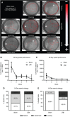
Clinical studies suggest that traumatic brain injury (TBI) hastens cognitive decline and development of neuropathology resembling brain aging. Blood-brain barrier (BBB) disruption following TBI may contribute to the aging process by deregulating substance exchange between the brain and blood. We evaluated the effect of juvenile TBI (jTBI) on these processes by examining long-term alterations of BBB proteins, β-amyloid (Aβ) neuropathology, and cognitive changes. A controlled cortical impact was delivered to the parietal cortex of male rats at postnatal day 17, with behavioral studies and brain tissue evaluation at 60 days post-injury (dpi). Immunoglobulin G extravasation was unchanged, and jTBI animals had higher levels of tight-junction protein claudin 5 versus shams, suggesting the absence of BBB disruption. However, decreased P-glycoprotein (P-gp) on cortical blood vessels indicates modifications of BBB properties. In parallel, we observed higher levels of endogenous rodent Aβ in several brain regions of the jTBI group versus shams. In addition at 60 dpi, jTBI animals displayed systematic search strategies rather than relying on spatial memory during the water maze. Together, these alterations to the BBB phenotype after jTBI may contribute to the accumulation of toxic products, which in turn may induce cognitive differences and ultimately accelerate brain aging.

Figures

Figure 1
Changes in endothelial tight junctions 2 months after juvenile traumatic brain injury (jTBI). (A, B) Immunoglobulin G (IgG) extravasation using infrared immunolabeling showed higher staining intensity at the median eminence and glia limitans of both (A) sham and (B) jTBI. (C) IgG levels were unchanged as quantified in the bilateral parietal cortex and striatum, thus indicating an overall lack of overt blood–brain barrier (BBB) leakage. (D, E) Endothelial tight-junction protein claudin 5 (Cld5) was observed on endothelial cells of intraparenchymal vessels in both (D) sham and (E) jTBI and (F) Cld5 quantification in the parietal and temporal cortices shows that jTBI have significantly increased Cld5 staining (P<0.05), perhaps as a restorative mechanism to improve BBB function (*P<0.05; values are represented as mean±s.e.m.; IR, infrared; Cld5, claudin 5; white arrowhead in B=lesion cavity; scale bar in (D, E)=50 μm).
Figure 2
Juvenile traumatic brain injury (jTBI) changes proteins involved in cellular trafficking at the blood–brain barrier (BBB). (A, B) Lipoprotein-related receptor protein 1 (LRP1) immunostaining is demonstrated in both the vascular walls (white arrowheads in D, E) as well as neuronal compartments (white arrows in A, B) in the cortex of a representative sham. (C) Quantification of LRP1 immunostaining in the parietal and temporal cortex shows that jTBI animals have lower overall staining of LRP1, but no significant differences were found between groups. (D, E) P-glycoprotein (P-gp) immunostaining is specific for endothelial cells (green), as shown in close proximity to the end-feet of glial fibrillary acidic protein (GFAP)-positive astrocytes (red) in both (D) sham and (E) jTBI. (F) P-gp quantification in the parietal and temporal cortices shows a significant decrease in vascular P-gp transporter in jTBI compared with sham (P<0.05). (G, H) Protein levels of jTBI are also significantly lower than sham, as shown in representative cases and quantification (P<0.05) (*P<0.05; values are represented as mean±s.e.m.; scale bar in (A)=20 μm; (B, D, E)=50 μm).
Figure 3
Immunoreactivity patterns with a rodent-Aβ antibody. (AH) Positive staining is detected in several brain regions following formic acid pretreatment and classical immunostaining using the specific rodent-Aβ antibody, as shown in representative sections from juvenile traumatic brain injury (jTBI) animals. (A) Temporal cortex shows specificity of the rod-Aβ antibody (labeled with goat anti-mouse λ488 nm), with negative staining using the secondary antibody alone on the same section (second alone, preincubation protocol with goat anti-mouse λ594 nm). (B) Western blotting from the parietal/temporal cortex shows the rod-Aβ antibody has high specificity for rat cortex, but not for human Aβ1-40 or Aβ1-42 peptides, while the 6E10 antibody has high specificity for human Aβ peptides at 4 kDa and larger aggregates, but no signal for rat tissue. In the rat, the rod-Aβ antibody reveals a prominent β-amyloid precursor protein (APP) band and several smaller fragments, indicating positive immunoreactivity for both Aβ aggregates or APP fragments. (C) In an example from the frontal cortex, rod-Aβ stains a cluster of several extracellular diffuse deposits (white arrows) and vascular labeling (white arrowheads) near the molecular and superficial cortical layers. (D) In an example from the parietal cortex, rod-Aβ staining is often surrounded by IBA1-positive microglial processes with an abnormal morphology (white arrowheads) suggestive of an immune response. (EH) Coincubation with both rod-Aβ antibody (raised in mouse, secondary λ488 nm) and a C-terminal antibody against Aβ42 (raised in rabbit, secondary λ594 nm) in the frontal cortex, shows several areas of colocalization (white arrowheads) as well as areas without overlap, indicating the presence of several Aβ species (Aβ, β-amyloid; rod-Aβ, rodent β-amyloid antibody; second alone, secondary antibody alone; M, marker; IBA1, ionized calcium binding adapter molecule 1; scale bars in (A, C)=100 μm; (D)=40 μm; (E, F, G, H)=30 μm).
Figure 4
Anterior to posterior patterns of rodent-Aβ distribution. (A) Representative coronal sections with outlines of rodent-Aβ positive staining in juvenile traumatic brain injury (jTBI) are shown at bregma +2.0 mm, −0.4, −1.4, and −5.2 mm. The jTBI lesion cavity is apparent in the parietal cortex at −1.4 mm and still visible at bregma −0.4 and −3.8 mm to a lesser extent (black arrowheads). (B) All positive rodent-Aβ immunoreactivity was quantified to obtain %Aβ load relative to total brain area at 8 bregma levels, with significant changes along the anterior-to-posterior axis (P<0.042), but no significant changes between groups at any single level. (C) Summation of total brain Aβ load was higher in jTBI versus sham (P<0.073), with jTBI animals showing high staining variability common in models of natural Aβ accumulation (values are represented as mean±s.e.m.; Aβ, β-amyloid).
Figure 5
Figure 5
Differences in water maze strategies in adulthood. (A) Eight swim patterns/learning strategies (red lines) were identified and divided into three categories from the most (+) to the least (−) efficient: spatial, systematic, and looping. (B, C) Water maze performance is unchanged between groups at (B) 30 days and (C) 60 days post-juvenile traumatic brain injury (jTBI); however, both groups improve and learn the task within each time point (block 1 to 5) and between testing time points (comparison of block 1 at 30 versus 60 days). (D) Swim strategy use was evenly distributed among sham and jTBI groups at 30 days, but not at 60 days. (E) By 60 days, jTBI animals used strategies similar to those used at 30 days (primarily systematic and looping patterns), but sham controls relied on significantly more spatial memory strategies compared with jTBI and to their prior performance at 30 days (P<0.05), (values are represented as mean±s.e.m.; jTBI, juvenile traumatic brain injury; +and − in A=most and least efficient, respectively). The color reproduction of this figure is available on the Journal of Cerebral Blood Flow & Metabolism journal online.
Figure 6
Summary of parallel changes in adulthood after early brain injury. A schematic representation of events 2 months after juvenile traumatic brain injury (jTBI) shows phenotypic alterations to the blood–brain barrier (BBB) changes occurring in parallel with molecular and behavioral dysfunction. Specifically, lack of immunoglobulin G (IgG) extravasation from the blood into the tissue and increased levels of tight-junction marker claudin 5 (cld5) indicate an intact BBB during adulthood. However, in an environment with decreased levels of P-glycoprotein (P-gp), and unchanged levels of lipoprotein-related receptor protein 1 (LRP1) and receptor for advanced glycation end products (RAGE), proper metabolism of toxic proteins such as Aβ can be hindered. As a result, the BBB phenotype impairs normal clearance of rod-Aβ and may promote its abnormal accumulation inside the brain in the parenchyma and vascular walls (adapted from Andras et al.).

Similar articles

See all similar articles

Cited by 30 articles

See all "Cited by" articles

Publication types

MeSH terms

Feedback