Habitual short sleep impacts frontal switch mechanism in attention to novelty
- PMID: 22131603
- PMCID: PMC3208843
- DOI: 10.5665/sleep.1430
Habitual short sleep impacts frontal switch mechanism in attention to novelty
Abstract
Study objectives: Reduced time in bed relative to biological sleep need is common. The impact of habitual short sleep on auditory attention has not been studied to date. In the current study, we utilized novelty oddball tasks to evaluate the effect of habitual short sleep on brain function underlying attention control processes measured by the mismatch negativity (MMN, index of pre-attentive stage), P3a (attention-dependent), and P3b (memory-dependent) event related brain potentials (ERPs). An extended time in bed in a separate study was used to evaluate the possible reversal of the impairments of these processes in habitual short sleepers.
Methods: Ten self-defined short sleepers (total sleep time [TST] ≤ 6 h) and 9 normal-sleeping subjects with TST 7-8 h, participated. ERPs were recorded via a 64-channel EEG system. Two test conditions: "ignore" and "attend" were implemented. The ERPs were analyzed and compared between groups on the 2 task conditions and frontal/central/parietal electrodes by 3-factor ANOVA. Sleep diary data were compared between groups by t-test. Sleep was recorded by the Zeo sleep monitoring system for a week in both habitual and extended sleep conditions at home.
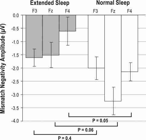
Results: The main findings of the present study show that short sleeping individuals had deficiency in activity of the MMN and P3a brain responses over frontal areas compared to normal-sleeping subjects. The P3b amplitude was increased over frontal areas and decreased over parietal with respect to the control group. Extension of time in bed for one week increased TST (from 5.7 h to 7.4 h), and concomitantly MMN amplitude increased from -0.1 μV up to -1.25 μV over frontal areas.
Conclusions: Reduced time in bed is associated with deficiency of the neuronal process associated with change detection, which may recover after one week of sleep extension, whereas attention-dependent neural processes do not normalize after this period of time in habitually short sleeping individuals and may require longer recovery periods.
Keywords: ERPs; Short sleep; attention; memory; sleep extension.
Figures
Similar articles
-
Connectivity analysis of novelty process in habitual short sleepers.Neuroimage. 2012 Nov 15;63(3):1001-10. doi: 10.1016/j.neuroimage.2012.08.011. Epub 2012 Aug 11. Neuroimage. 2012. PMID: 22906789
-
Sleep extension normalizes ERP of waking auditory sensory gating in healthy habitually short sleeping individuals.PLoS One. 2013;8(3):e59007. doi: 10.1371/journal.pone.0059007. Epub 2013 Mar 8. PLoS One. 2013. PMID: 23520548 Free PMC article. Clinical Trial.
-
The effects of early and late night partial sleep deprivation on automatic and selective attention: An ERP study.Brain Res. 2010 Jan 13;1308:87-99. doi: 10.1016/j.brainres.2009.09.090. Epub 2009 Sep 30. Brain Res. 2010. PMID: 19799884
-
ERPs studies of cognitive processing during sleep.Int J Psychol. 2009 Aug;44(4):290-304. doi: 10.1080/00207590802194234. Int J Psychol. 2009. PMID: 22029558 Review.
-
[Study of sensory memory reflected by mismatch negativity and its clinical application].Seishin Shinkeigaku Zasshi. 2004;106(1):1-16. Seishin Shinkeigaku Zasshi. 2004. PMID: 15049124 Review. Japanese.
Cited by 13 articles
-
Performance of an Ambulatory Dry-EEG Device for Auditory Closed-Loop Stimulation of Sleep Slow Oscillations in the Home Environment.Front Hum Neurosci. 2018 Mar 8;12:88. doi: 10.3389/fnhum.2018.00088. eCollection 2018. Front Hum Neurosci. 2018. PMID: 29568267 Free PMC article.
-
A single night of sleep loss impairs objective but not subjective working memory performance in a sex-dependent manner.J Sleep Res. 2019 Feb;28(1):e12651. doi: 10.1111/jsr.12651. Epub 2018 Jan 31. J Sleep Res. 2019. PMID: 29383809 Free PMC article.
-
The sleep-deprived human brain.Nat Rev Neurosci. 2017 Jul;18(7):404-418. doi: 10.1038/nrn.2017.55. Epub 2017 May 18. Nat Rev Neurosci. 2017. PMID: 28515433 Free PMC article. Review.
-
Variability in Cumulative Habitual Sleep Duration Predicts Waking Functional Connectivity.Sleep. 2016 Jan 1;39(1):87-95. doi: 10.5665/sleep.5324. Sleep. 2016. PMID: 26414900 Free PMC article.
-
Performance of a Portable Sleep Monitoring Device in Individuals with High Versus Low Sleep Efficiency.J Clin Sleep Med. 2016 Jan;12(1):95-103. doi: 10.5664/jcsm.5404. J Clin Sleep Med. 2016. PMID: 26285110 Free PMC article.