Review
. 2011 Nov 3;12(12):723-38.
doi: 10.1038/nrn3114.

Neurovascular pathways to neurodegeneration in Alzheimer's disease and other disorders

Affiliations
Free PMC article
Review

Neurovascular pathways to neurodegeneration in Alzheimer's disease and other disorders

Berislav V Zlokovic. Nat Rev Neurosci. .
Free PMC article

Abstract

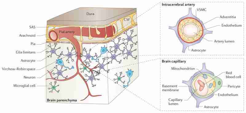
The neurovascular unit (NVU) comprises brain endothelial cells, pericytes or vascular smooth muscle cells, glia and neurons. The NVU controls blood-brain barrier (BBB) permeability and cerebral blood flow, and maintains the chemical composition of the neuronal 'milieu', which is required for proper functioning of neuronal circuits. Recent evidence indicates that BBB dysfunction is associated with the accumulation of several vasculotoxic and neurotoxic molecules within brain parenchyma, a reduction in cerebral blood flow, and hypoxia. Together, these vascular-derived insults might initiate and/or contribute to neuronal degeneration. This article examines mechanisms of BBB dysfunction in neurodegenerative disorders, notably Alzheimer's disease, and highlights therapeutic opportunities relating to these neurovascular deficits.

Conflict of interest statement

Competing interests statement

The author declares competing financial interests: see web version for details.

Figures

Box 1
The two-hit vascular hypothesis for Alzheimer’s disease
Box 2
A model of multiple-target, multiple-action neurovascular medicine
Figure 1. Cerebral microcirculation and the neurovascular unit
In the brain, pial arteries run through the subarachnoid space (SAS), which contains the cerebrospinal fluid (CSF). These vessels give rise to intracerebral arteries, which penetrate into brain parenchyma. Intracerebral arteries are separated from brain parenchyma by a single, interrupted layer of elongated fibroblast-like cells of the pia and the astrocyte-derived glia limitans membrane that forms the outer wall of the perivascular Virchow–Robin space. These arteries branch into smaller arteries and subsequently arterioles, which lose support from the glia limitans and give rise to pre-capillary arterioles and brain capillaries. In an intracerebral artery, the vascular smooth muscle cell (VSMC) layer occupies most of the vessel wall. At the brain capillary level, vascular endothelial cells and pericytes are attached to the basement membrane. Pericyte processes encase most of the capillary wall, and they communicate with endothelial cells directly through synapse-like contacts containing connexins and N-cadherin. Astrocyte end-foot processes encase the capillary wall, which is composed of endothelium and pericytes. Resting microglia have a ‘ramified’ shape and can sense neuronal injury.
Figure 2. Blood–brain barrier transport mechanisms
Small lipophilic drugs, oxygen and carbon dioxide diffuse across the blood–brain barrier (BBB), whereas ions require ATP-dependent transporters such as the (Na++K+) ATPase. Transporters for nutrients include the glucose transporter 1 (GLUT1; also known as solute carrier family 2, facilitated glucose transporter member 1 (SLC2A1)), the lactate transporter monocarboxylate transporter 1 (MCT1) and the L1 and y+ transporters for large neutral and cationic essential amino acids, respectively. These four transporters are expressed at both the luminal and albuminal membranes. Non-essential amino acid transporters (the alanine, serine and cysteine preferring system (ASC), and the alanine preferring system (A)) and excitatory amino acid transporter 1 (EAAT1), EAAT2 and EAAT3 are located at the abluminal side. The ATP-binding cassette (ABC) efflux transporters that are found in the endothelial cells include multidrug resistance protein 1 (ABCB1; also known as ATP-binding cassette subfamily B member 1) and solute carrier organic anion transporter family member 1C1 (OATP1C1). Finally, transporters for peptides or proteins include the endothelial protein C receptor (EPCR) for activated protein C (APC); the insulin receptors (IRs) and the transferrin receptors (TFRs), which are associated with caveolin 1 (CAV1); low-density lipoprotein receptor-related protein 1 (LRP1) for amyloid-β, peptide transport system 1 (PTS1) for encephalins; and the PTS2 and PTS4–vasopressin V1a receptor (V1AR) for arginine vasopressin.
Figure 3. Vascular-mediated neuronal damage and neurodegeneration
a, Blood–brain barrier (BBB) breakdown that is caused by pericyte detachment leads to leakage of serum proteins and focal microhaemorrhages, with extravasation of red blood cells (RBCs). RBCs release haemoglobin, which is a source of iron. In turn, this metal catalyses the formation of toxic reactive oxygen species (ROS) that mediate neuronal injury. Albumin promotes the development of vasogenic oedema, contributing to hypoperfusion and hypoxia of the nervous tissue, which aggravates neuronal injury. A defective BBB allows several potentially vasculotoxic and neurotoxic proteins (for example, thrombin, fibrin and plasmin) to enter the brain. b, Progressive reductions in cerebral blood flow (CBF) lead to increasing neuronal dysfunction. Mild hypoperfusion, oligaemia, leads to a decrease in protein synthesis, whereas more-severe reductions in CBF, leading to hypoxia, cause an array of detrimental effects.
Figure 4. The role of blood–brain barrier transport in brain homeostasis of amyloid-β
Amyloid-β (Aβ) is produced from the amyloid-β precursor protein (APP), both in the brain and in peripheral tissues. Clearance of amyloid-β from the brain normally maintains its low levels in the brain. This peptide is cleared across the blood– brain barrier (BBB) by the low-density lipoprotein receptor-related protein 1 (LRP1). LRP1 mediates rapid efflux of a free, unbound form of amyloid-β and of amyloid-β bound to apolipoprotein E2 (APOE2), APOE3 or α2-macroglobulin (not shown) from the brain’s interstitial fluid into the blood, and APOE4 inhibits such transport. LRP2 eliminates amyloid-β that is bound to clusterin (CLU; also known as apolipoprotein J (APOJ)) by transport across the BBB, and shows a preference for the 42-aminoacid form of this peptide. ATP-binding cassette subfamily A member 1 (ABCA1; also known as cholesterol efflux regulatory protein) mediates amyloid-β efflux from the brain endothelium to blood across the luminal side of the BBB (not shown). Cerebral endothelial cells, pericytes, vascular smooth muscle cells, astrocytes, microglia and neurons express different amyloid-β-degrading enzymes, including neprilysin (NEP), insulin-degrading enzyme (IDE), tissue plasminogen activator (tPA) and matrix metalloproteinases (MMPs), which contribute to amyloid-β clearance. In the circulation, amyloid-β is bound mainly to soluble LRP1 (sLRP1), which normally prevents its entry into the brain. Systemic clearance of amyloid-β is mediated by its removal by the liver and kidneys. The receptor for advanced glycation end products (RAGE) provides the key mechanism for influx of peripheral amyloid-β into the brain across the BBB either as a free, unbound plasma-derived peptide and/or by amyloid-β-laden monocytes. Faulty vascular clearance of amyloid-β from the brain and/or an increased re-entry of peripheral amyloid-β across the blood vessels into the brain can elevate amyloid-β levels in the brain parenchyma and around cerebral blood vessels. At pathophysiological concentrations, amyloid-β forms neurotoxic oligomers and also self-aggregates, which leads to the development of cerebral β-amyloidosis and cerebral amyloid angiopathy.
Figure 5. A model of vascular damage in Alzheimer’s disease
a, In the early stages of Alzheimer’s disease, small pial and intracerebral arteries develop a hypercontractile phenotype that underlies dysregulated cerebral blood flow (CBF). This phenotype is accompanied by diminished amyloid-β clearance by the vascular smooth muscle cells (VSMCs). In the later phases of Alzheimer’s disease, amyloid deposition in the walls of intracerebral arteries leads to cerebral amyloid angiopathy (CAA), pronounced reductions in CBF, atrophy of the VSMC layer and rupture of the vessels causing microbleeds. b, At the level of capillaries in the early stages of Alzheimer’s disease, blood–brain barrier (BBB) dysfunction leads to a faulty amyloid-β clearance and accumulation of neurotoxic amyloid-β oligomers in the interstitial fluid (ISF), microhaemorrhages and accumulation of toxic blood-derived molecules (that is, thrombin and fibrin), which affect synaptic and neuronal function. Hyperphosphorylated tau (p-tau) accumulates in neurons in response to hypoperfusion and/or rising amyloid-β levels. At this point, microglia begin to sense neuronal injury. In the later stages of the disease in brain capillaries, microvascular degeneration leads to increased deposition of basement membrane proteins and perivascular amyloid. The deposited proteins and amyloid obstruct capillary blood flow, resulting in failure of the efflux pumps, accumulation of metabolic waste products, changes in pH and electrolyte composition and, subsequently, synaptic and neuronal dysfunction. Neurofibrillary tangles (NFTs) accumulate in response to ischaemic injury and rising amyloid-β levels. Activation of microglia and astrocytes is associated with a pronounced inflammatory response. ROS, reactive oxygen species.

Similar articles

See all similar articles

Cited by 709 articles

See all "Cited by" articles

Publication types

MeSH terms

Feedback