. 2011 Apr 29;286(17):15317-31.
doi: 10.1074/jbc.M110.209296. Epub 2011 Mar 3.

Seeding of normal Tau by pathological Tau conformers drives pathogenesis of Alzheimer-like tangles

Affiliations
Free PMC article

Seeding of normal Tau by pathological Tau conformers drives pathogenesis of Alzheimer-like tangles

Jing L Guo et al. J Biol Chem. .
Free PMC article

Abstract

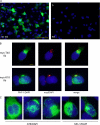
Neurofibrillary tangles (NFTs) in Alzheimer disease and related tauopathies are composed of insoluble hyperphosphorylated Tau protein, but the mechanisms underlying the conversion of highly soluble Tau into insoluble NFTs remain elusive. Here, we demonstrate that introduction of minute quantities of misfolded preformed Tau fibrils (Tau pffs) into Tau-expressing cells rapidly recruit large amounts of soluble Tau into filamentous inclusions resembling NFTs with unprecedented efficiency, suggesting a "seeding"-recruitment process as a highly plausible mechanism underlying NFT formation in vivo. Consistent with the emerging concept of prion-like transmissibility of disease-causing amyloidogenic proteins, we found that spontaneous uptake of Tau pffs into cells is likely mediated by endocytosis, suggesting a potential mechanism for the propagation of Tau lesions in tauopathy brains. Furthermore, sequestration of soluble Tau by pff-induced Tau aggregates attenuates microtubule overstabilization in Tau-expressing cells, supporting the hypothesis of a Tau loss-of-function toxicity in cells harboring NFTs. In summary, our study establishes a cellular system that robustly develops authentic NFT-like Tau aggregates, which provides mechanistic insights into NFT pathogenesis and a potential tool for identifying Tau-based therapeutics.

Figures

FIGURE 1.
FIGURE 1.
Tau pffs seed endogenous Tau fibrillization in WT-T40-transfected QBI-293 cells. A, phosphorylated Tau recognized by phospho-Tau antibody PHF-1 (green) was completely soluble in WT-T40-transfected cells treated with BioPORTER reagent alone. Soluble proteins were extracted (ext) by 1% Triton X-100 during fixing of the cells in panel b but not in panel a. B, intracellular accumulation of insoluble Tau recognized by PHF-1 can be induced by both Myc-T40 and Myc-K18 pffs (referred as myc-T40 fib and myc-K18 fib) transduced using BioPORTER reagent. Exogenous pffs were immunostained with polyclonal anti-Myc antibody (red). C, induced Tau aggregates showed a range of morphologies that can be recognized by phospho-Tau antibody AT8 and conformational-dependent antibody MC-1. B and C, cells were extracted with 1% Triton X-100 during fixing. In all panels, cell nuclei were stained by DAPI (blue). Magnification: ×20 for A; ×40 for B and C. Scale bars, 100 μm for A; 10 μm for B and C.
FIGURE 2.
P301L mutation enhances pff-induced aggregation. A, QBI-293 cells were transiently transfected with T40 with P301L mutation (T40/P301L) and transduced with Myc-K18 fibrils (fib) (panel a) or Myc-K18/P301L fibrils (panel b) using BioPORTER reagent. Abundant Tau aggregates were detected using mAb PHF-1 (green) after simultaneous 1% Triton X-100 extraction during fixation to remove soluble proteins. B, sequential extraction was performed on T40/P301L-transfected cells treated with BioPORTER reagent alone (control), with Myc-K18 fibrils (K18 fib), or Myc-K18/P301L fibrils (K18/PL fib). T indicates the cellular fraction recovered in 1% Triton X-100 lysis buffer. S indicates the Triton-insoluble fraction solubilized in 1% SDS lysis buffer. Equal proportions of Triton and SDS fractions were loaded on SDS-polyacrylamide gels. Immunoblotting with polyclonal Tau Ab 17025 and PHF-1 revealed accumulation of abundant Triton-insoluble Tau in fibril transduced cells. GAPDH served as a loading control. C, sandwich Tau ELISA on cell lysates from four independent experiments (exp) confirmed induction of significant amount of Triton-insoluble Tau with fibril transduction, which was always accompanied by a reduction in soluble Tau. Magnification: ×40. Scale bar, 50 μm.
FIGURE 3.
Internalization of small quantities of Tau pffs is sufficient to induce robust Tau aggregation. QBI-293 cells were transiently transfected with mutant T40/P301L Tau and transduced with Myc-K18/P301L pffs using BioPORTER reagent. A, incubation of fibril-transduced cells with polyclonal anti-Myc antibody before fixing (live, red) followed by staining of fixed and permeabilized cells with anti-Myc mAb 9E10 (fixed, green) showed extensive colocalization, suggesting the majority of the pffs detected were merely extracellularly associated with cell membranes. Arrows point to truly internalized fibrils that were only recognized by 9E10 applied to fixed and permeabilized cells but not polyclonal anti-Myc antibody used on live cells. B, double labeling of phospho-aggregates by PHF-1 (green) and exogenous pffs by polyclonal anti-Myc (red) showed very little colocalization. Arrows point to aggregates with colocalizing pff seeds. Aggregates without arrows do not contain obvious pff seeds. Magnification: ×20. Scale bar, 100 μm.
FIGURE 4.
pff-induced Tau aggregates consist of filamentous structures. T40/P301L-transfected QBI-293 cells were transduced with Myc-K18/P301L fibrils using BioPORTER reagent. A, immuno-EM using mAb PHF-1 showed abundant filamentous Tau aggregates in the cytoplasm. Arrows indicate mitochondria and/or vesicular structures in close association with Tau fibrils. B, routine EM showing pff-induced Tau fibrils. Magnification: ×12,000 for A, panel a; ×30,000 for A, panel b; 50,000 for B. Scale bars: 2 μm for A, panel a; 500 nm for A, panel b, and B.
FIGURE 5.
Time-dependent development of insoluble Tau aggregates. QBI-293 cells transfected with T40/P301L mutant Tau were transduced with Myc-K18/P301L fibrils using BioPORTER reagent. A, at t = 3 h after the addition of fibril-reagent complex, a small percentage of cells started showing accumulations of insoluble Tau (PHF-1) with focal inclusions colocalizing with pff staining (myc). B, at t = 6 h, more cells developed aggregates, most of which are skein-like and diffusely distributed throughout the cytoplasm. A small population of cells showed ThS-positive large aggregates. C, large aggregates recognized by ThS were more frequently seen at t = 24 h after fibril addition. Soluble proteins were extracted by 1% Triton X-100 during fixing for all. Magnification: ×40. Scale bar, 50 μm.
FIGURE 6.
Newly synthesized Tau is rapidly recruited into insoluble aggregates. One day after Myc-K18/P301L fibril transduction on QBI-293 cells transiently transfected with T40/P301L, cells were pulsed with [35S]methionine for 20 min and chased for different durations (0–6 h). Cell lysates were sequentially extracted using 1% Triton X-100 followed by 1% SDS lysis buffer, and Tau was immunoprecipitated from both fractions with mAbs T46 and Tau 5. Equal proportions of Triton (T) and SDS (S) fractions were loaded on SDS-PAGE for autoradiography. A, autoradiograph indicating that newly synthesized Tau remained soluble in cells treated with reagent alone (Rg control) but rapidly turned insoluble in fibril-transduced cells (Fib Td). B, quantification from two independent experiments showing the distribution of radiolabeled Tau in the soluble and insoluble fractions over time (error bar, standard error).
FIGURE 7.
Tau aggregation results in reduced MT stability. A, QBI-293 cells transfected with T40/P301L and transduced with Myc-K18 (K18 fib) or Myc-K18/P301L (K18/PL fib) pffs showed significant reductions in MT bundling compared with control cells treated with BioPORTER transduction reagent alone (control), as revealed by acetylated tubulin (Ac-tub) staining. B, double staining of PHF-1 and Ac-tub showed that cells with MT bundles usually showed diffuse Tau immunostaining (arrows), whereas those with Tau aggregates often lacked bundling (arrowheads). C, quantification from three independent experiments demonstrated statistically significant reduction in the percentage of cells with MT bundles in the presence of fibril transduction (error bar, standard error; *, p < 0.05). D, Ac-tub sandwich ELISA on samples from four independent experiments showed significant decrease in Ac-tub levels associated with fibril transduction (error bar, standard error; *, p < 0.05). E and F, Ac-tub and Tau sandwich ELISA revealed highly significant correlation of Ac-tub levels with Triton-soluble Tau (E, p < 0.0005), but not with Triton-insoluble Tau (F, p > 0.05), across control and fibril-transduced samples from four independent experiments. Magnification: ×20 for A; ×40 for B. Scale bar, 100 μm for A; 50 μm for B.
FIGURE 8.
Spontaneous uptake of pffs without transduction reagent also induces intracellular Tau aggregation. A, T40/P301L aggregation induced by Myc-K18/P301L pffs alone after 30 h of incubation at 37 °C (panel a) or 4 h incubation at 37 °C (panel b). Minimal aggregation was observed with 4 h of incubation at 4 °C (panel c). For all conditions, cells were trypsinized and replated after the designated incubation period and fixed 48 h after the initial addition of fibrils. Soluble proteins were extracted with 1% Triton X-100 during fixing. White, PHF-1. B, 48 h incubation with Myc-K18/P301L pffs at 37 °C led to significant accumulation of Triton-insoluble T40/P301L detected on immunoblots. Control cells were transfected with T40/P301L without fibril transduction. C, 4 h of incubation with Myc-K18/P301L fibrils at 37 °C but not at 4 °C resulted in detectable T40/P301L Triton-insoluble aggregates. D, quantification from four independent experiments showed significant reduction in aggregate-bearing cells when a 4-h fibril incubation was performed at 4 °C instead of 37 °C (error bar, standard error; *, p < 0.05 compared with 4 h of incubation at 37 °C). Thirty-hour incubation with Tau pffs at 37 °C resulted in the highest incidence of aggregates (n = 3; error bar, standard error; **, p < 0.01 compared with 4 h incubation at 37 °C). E, Myc-tagged pffs were barely visible in cells transduced with fibrils (fib) alone without BioPORTER reagent. White, anti-Myc Ab. F, exogenous Myc-tagged Tau pffs were nondetectable with anti-Myc mAb 9E10 via immunoblot under various transduction paradigms: transduction of pffs with BioPORTER reagent (fib Td with Rg), 30 h of incubation with pffs alone (30 h fib alone), or 4 h incubation with fibrils alone (4 h fib alone). Cells were trypsinized and replated to new wells after the designated incubation time so that the majority of extracellularly associated pffs were removed. Total fib input, amount of Tau pffs used for transduction was added to cells immediately before lysing. *, nonspecific bands. B, C, and F, T indicates 1% Triton X-100 fraction; S indicates 1% SDS fraction. Equal proportions of Triton and SDS fractions were loaded on SDS-polyacrylamide gels. Magnification: ×20. Scale bar, 100 μm.
FIGURE 9.
WGA promotes pff-induced Tau aggregation by enhancing spontaneous pff uptake. A, T40/P301L-transfected cells were incubated with Myc-K18/P301L pffs in the presence of 0, 5, 10, and 15 μg/ml of WGA (0, 5, 10, and 15) without BioPORTER reagent. Increasing doses of WGA resulted in more frequent phospho-Tau aggregates. Green, PHF-1 with 1% Triton X-100 added during fixing of the cells. B, quantification from three independent experiments indicated that WGA dose-dependently increased the percentage of cells with aggregation (error bar, standard error; *, p < 0.05; **, p < 0.01). C, WGA treatment increased accumulation of Triton-insoluble Tau shown on immunoblots. T indicates 1% Triton X-100 fraction; S indicates 1% SDS fraction. Equal proportions of Triton X-100 and SDS fractions were loaded on SDS-PAGE. D, WGA also dose-dependently (0, 5, 10, and 15 μg/ml) increased the amount of cell-associated pffs revealed by Myc immunoreactivities (red). E, two-stage immunostaining (see “Experimental Procedures”) performed on cells treated with 15 μg/ml of WGA showed frequent occurrence of truly internalized pffs that were only labeled by 9E10 (green, fixed) applied to fixed and permeabilized cells but not polyclonal anti-Myc antibody incubated with live cells (red, live). Arrows point to examples of truly intracellular pffs. F, GlcNAc, which inhibits binding of WGA to plasma membrane, dramatically attenuated the aggregation-enhancing effects of WGA. G, quantification from three independent experiments showed significant reduction of Tau aggregation in WGA-treated fibril transduced cells in the presence of GlcNAc (error bar, standard error; *, p < 0.05). Magnification: ×20 in A and F; ×40 in D and E. Scale bars, 100 μm in A and F; 50 μm in D and E.

Similar articles

See all similar articles

Cited by 224 articles

See all "Cited by" articles

Publication types

Feedback