Sulforaphane, a dietary component of broccoli/broccoli sprouts, inhibits breast cancer stem cells
- PMID: 20388854
- PMCID: PMC2862133
- DOI: 10.1158/1078-0432.CCR-09-2937
Sulforaphane, a dietary component of broccoli/broccoli sprouts, inhibits breast cancer stem cells
Abstract
Purpose: The existence of cancer stem cells (CSCs) in breast cancer has profound implications for cancer prevention. In this study, we evaluated sulforaphane, a natural compound derived from broccoli/broccoli sprouts, for its efficacy to inhibit breast CSCs and its potential mechanism.
Experimental design: Aldefluor assay and mammosphere formation assay were used to evaluate the effect of sulforaphane on breast CSCs in vitro. A nonobese diabetic/severe combined immunodeficient xenograft model was used to determine whether sulforaphane could target breast CSCs in vivo, as assessed by Aldefluor assay, and tumor growth upon cell reimplantation in secondary mice. The potential mechanism was investigated using Western blotting analysis and beta-catenin reporter assay.
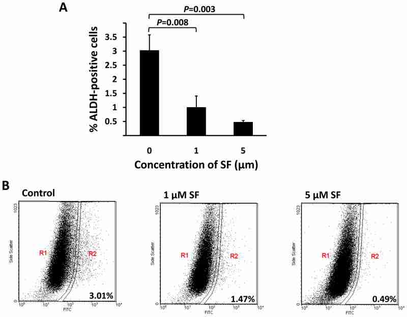
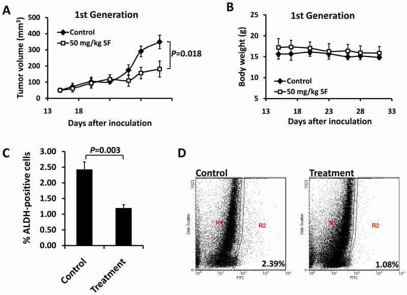
Results: Sulforaphane (1-5 micromol/L) decreased aldehyde dehydrogenase-positive cell population by 65% to 80% in human breast cancer cells (P < 0.01) and reduced the size and number of primary mammospheres by 8- to 125-fold and 45% to 75% (P < 0.01), respectively. Daily injection with 50 mg/kg sulforaphane for 2 weeks reduced aldehyde dehydrogenase-positive cells by >50% in nonobese diabetic/severe combined immunodeficient xenograft tumors (P = 0.003). Sulforaphane eliminated breast CSCs in vivo, thereby abrogating tumor growth after the reimplantation of primary tumor cells into the secondary mice (P < 0.01). Western blotting analysis and beta-catenin reporter assay showed that sulforaphane downregulated the Wnt/beta-catenin self-renewal pathway.
Conclusions: Sulforaphane inhibits breast CSCs and downregulates the Wnt/beta-catenin self-renewal pathway. These findings support the use of sulforaphane for the chemoprevention of breast cancer stem cells and warrant further clinical evaluation.
Copyright 2010 AACR.
Figures
Similar articles
-
Targeting cancer stem cells with sulforaphane, a dietary component from broccoli and broccoli sprouts.Future Oncol. 2013 Aug;9(8):1097-103. doi: 10.2217/fon.13.108. Future Oncol. 2013. PMID: 23902242
-
Synergistic activity of sorafenib and sulforaphane abolishes pancreatic cancer stem cell characteristics.Cancer Res. 2010 Jun 15;70(12):5004-13. doi: 10.1158/0008-5472.CAN-10-0066. Epub 2010 Jun 8. Cancer Res. 2010. PMID: 20530687
-
Sulforaphane Suppresses the Growth of Triple-negative Breast Cancer Stem-like Cells In vitro and In vivo.Cancer Prev Res (Phila). 2019 Mar;12(3):147-158. doi: 10.1158/1940-6207.CAPR-18-0241. Epub 2019 Jan 24. Cancer Prev Res (Phila). 2019. PMID: 30679159 Free PMC article.
-
Implications of cancer stem cell theory for cancer chemoprevention by natural dietary compounds.J Nutr Biochem. 2011 Sep;22(9):799-806. doi: 10.1016/j.jnutbio.2010.11.001. Epub 2011 Feb 4. J Nutr Biochem. 2011. PMID: 21295962 Free PMC article. Review.
-
Phytochemicals as Innovative Therapeutic Tools against Cancer Stem Cells.Int J Mol Sci. 2015 Jul 10;16(7):15727-42. doi: 10.3390/ijms160715727. Int J Mol Sci. 2015. PMID: 26184171 Free PMC article. Review.
Cited by 167 articles
-
Targeting STAT3 signaling using stabilised sulforaphane (SFX-01) inhibits endocrine resistant stem-like cells in ER-positive breast cancer.Oncogene. 2020 Jun;39(25):4896-4908. doi: 10.1038/s41388-020-1335-z. Epub 2020 May 30. Oncogene. 2020. PMID: 32472077 Free PMC article.
-
Breast Cancer Prevention-Is there a Future for Sulforaphane and Its Analogs?Nutrients. 2020 May 27;12(6):1559. doi: 10.3390/nu12061559. Nutrients. 2020. PMID: 32471217 Free PMC article. Review.
-
The Role of Secondary Metabolites on Gynecologic Cancer Therapy: Some Pathways and Mechanisms.Turk J Pharm Sci. 2017 Dec;14(3):324-334. doi: 10.4274/tjps.49368. Epub 2017 Nov 20. Turk J Pharm Sci. 2017. PMID: 32454632 Free PMC article. Review.
-
Sporadic activation of an oxidative stress-dependent NRF2-p53 signaling network in breast epithelial spheroids and premalignancies.Sci Signal. 2020 Apr 14;13(627):eaba4200. doi: 10.1126/scisignal.aba4200. Sci Signal. 2020. PMID: 32291314
-
Sulforaphene Suppresses Adipocyte Differentiation via Induction of Post-Translational Degradation of CCAAT/Enhancer Binding Protein Beta (C/EBPβ).Nutrients. 2020 Mar 13;12(3):758. doi: 10.3390/nu12030758. Nutrients. 2020. PMID: 32183002 Free PMC article.
Publication types
MeSH terms
Substances
Grant support
LinkOut - more resources
-
Full Text Sources
-
Other Literature Sources
-
Medical
-
Miscellaneous
