Profile and regulation of apolipoprotein E (ApoE) expression in the CNS in mice with targeting of green fluorescent protein gene to the ApoE locus
- PMID: 16687490
- PMCID: PMC6674234
- DOI: 10.1523/JNEUROSCI.5476-05.2006
Profile and regulation of apolipoprotein E (ApoE) expression in the CNS in mice with targeting of green fluorescent protein gene to the ApoE locus
Abstract
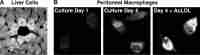
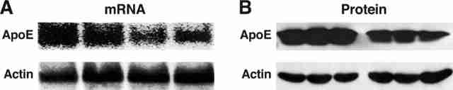
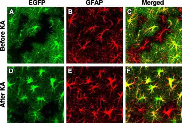
To study the profile and regulation of apolipoprotein E (apoE) expression in the CNS, we generated mice in which apoE expression can be detected in vivo with unprecedented sensitivity and resolution. cDNA encoding enhanced green fluorescent protein (EGFP) with a stop codon was inserted by gene targeting into the apoE gene locus (EGFPapoE) immediately after the translation initiation site. Insertion of EGFP into one apoE allele provides a real-time location marker of apoE expression in vivo; the remaining allele is sufficient to maintain normal cellular physiology. In heterozygous EGFPapoE mice, EGFP was highly expressed in hepatocytes and peritoneal macrophages. EGFP was also expressed in brain astrocytes; however some astrocytes (approximately 25%) expressed no EGFP, suggesting that a subset of these cells does not express apoE. EGFP was expressed in <10% of microglia after kainic acid treatment, suggesting that microglia are not a major source of brain apoE. Although hippocampal neurons did not express EGFP under normal conditions, kainic acid treatment induced intense expression of EGFP in injured neurons, demonstrating apoE expression in neurons in response to excitotoxic injury. The neuronal expression was confirmed by in situ hybridization of mouse apoE mRNA and by anti-apoE immunostaining. Smooth muscle cells of large blood vessels and cells surrounding small vessels in the CNS also strongly expressed EGFP, as did cells in the choroid plexus. EGFPapoE reporter mice will be useful for studying the regulation of apoE expression in the CNS and might provide insights into the diverse mechanisms of apoE4-related neurodegeneration.
Figures
Similar articles
-
Intron-3 retention/splicing controls neuronal expression of apolipoprotein E in the CNS.J Neurosci. 2008 Feb 6;28(6):1452-9. doi: 10.1523/JNEUROSCI.3253-07.2008. J Neurosci. 2008. PMID: 18256266 Free PMC article.
-
Presence of apolipoprotein E immunoreactivity in degenerating neurones of mice is dependent on the severity of kainic acid-induced lesion.Brain Res. 2000 Jun 23;868(2):165-75. doi: 10.1016/s0006-8993(00)02250-2. Brain Res. 2000. PMID: 10854569
-
Endocytosis of apoE-EGFP by primary human brain cultures.Cell Biol Int. 2002;26(9):761-70. doi: 10.1016/s1065-6995(02)90930-3. Cell Biol Int. 2002. PMID: 12377208
-
Astroglial regulation of apolipoprotein E expression in neuronal cells. Implications for Alzheimer's disease.J Biol Chem. 2004 Jan 30;279(5):3862-8. doi: 10.1074/jbc.M309475200. Epub 2003 Oct 29. J Biol Chem. 2004. PMID: 14585838
-
Apolipoprotein E as a target for developing new therapeutics for Alzheimer's disease based on studies from protein, RNA, and regulatory region of the gene.J Mol Neurosci. 2004;23(3):225-33. doi: 10.1385/JMN:23:3:225. J Mol Neurosci. 2004. PMID: 15181251 Review.
Cited by 147 articles
-
In Vivo Chimeric Alzheimer's Disease Modeling of Apolipoprotein E4 Toxicity in Human Neurons.Cell Rep. 2020 Jul 28;32(4):107962. doi: 10.1016/j.celrep.2020.107962. Cell Rep. 2020. PMID: 32726626 Free PMC article.
-
Apolipoprotein E expression pattern in human induced pluripotent stem cells during in vitro neural induction.F1000Res. 2020 May 12;9:353. doi: 10.12688/f1000research.23580.1. eCollection 2020. F1000Res. 2020. PMID: 32685135 Free PMC article.
-
Cell Type Impacts Accessibility of mRNA to Silencing by RNA Interference.Mol Ther Nucleic Acids. 2020 Jun 12;21:384-393. doi: 10.1016/j.omtn.2020.06.006. Online ahead of print. Mol Ther Nucleic Acids. 2020. PMID: 32650236 Free PMC article.
-
Choroid Plexus: The Orchestrator of Long-Range Signalling Within the CNS.Int J Mol Sci. 2020 Jul 4;21(13):4760. doi: 10.3390/ijms21134760. Int J Mol Sci. 2020. PMID: 32635478 Free PMC article. Review.
-
ABCA1/ApoE/HDL Signaling Pathway Facilitates Myelination and Oligodendrogenesis after Stroke.Int J Mol Sci. 2020 Jun 19;21(12):4369. doi: 10.3390/ijms21124369. Int J Mol Sci. 2020. PMID: 32575457 Free PMC article.
Publication types
MeSH terms
Substances
LinkOut - more resources
-
Full Text Sources
-
Other Literature Sources
-
Molecular Biology Databases
-
Miscellaneous
