Molecular mechanisms of cerebrospinal fluid production
- PMID: 15561411
- PMCID: PMC1890044
- DOI: 10.1016/j.neuroscience.2004.07.003
Molecular mechanisms of cerebrospinal fluid production
Abstract
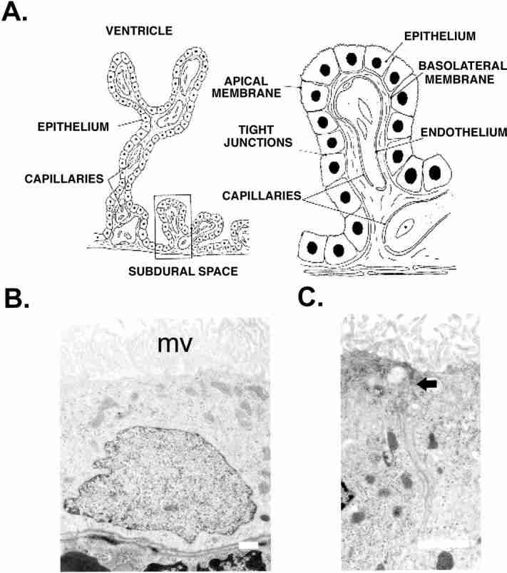
The epithelial cells of the choroid plexuses secrete cerebrospinal fluid (CSF), by a process which involves the transport of Na(+), Cl(-) and HCO(3)(-) from the blood to the ventricles of the brain. The unidirectional transport of ions is achieved due to the polarity of the epithelium, i.e. the ion transport proteins in the blood-facing (basolateral) membrane are different to those in the ventricular (apical) membrane. The movement of ions creates an osmotic gradient which drives the secretion of H(2)O. A variety of methods (e.g. isotope flux studies, electrophysiological, RT-PCR, in situ hybridization and immunocytochemistry) have been used to determine the expression of ion transporters and channels in the choroid plexus epithelium. Most of these transporters have now been localized to specific membranes. For example, Na(+)-K(+)ATPase, K(+) channels and Na(+)-2Cl(-)-K(+) cotransporters are expressed in the apical membrane. By contrast the basolateral membrane contains Cl(-)- HCO(3) exchangers, a variety of Na(+) coupled HCO(3)(-) transporters and K(+)-Cl(-) cotransporters. Aquaporin 1 mediates water transport at the apical membrane, but the route across the basolateral membrane is unknown. A model of CSF secretion by the mammalian choroid plexus is proposed which accommodates these proteins. The model also explains the mechanisms by which K(+) is transported from the CSF to the blood.
Figures
Similar articles
-
Mechanisms of CSF secretion by the choroid plexus.Microsc Res Tech. 2001 Jan 1;52(1):49-59. doi: 10.1002/1097-0029(20010101)52:1<49::AID-JEMT7>3.0.CO;2-C. Microsc Res Tech. 2001. PMID: 11135448 Review.
-
The murine choroid plexus epithelium expresses the 2Cl-/H+ exchanger ClC-7 and Na+/H+ exchanger NHE6 in the luminal membrane domain.Am J Physiol Cell Physiol. 2018 Apr 1;314(4):C439-C448. doi: 10.1152/ajpcell.00145.2017. Epub 2017 Dec 20. Am J Physiol Cell Physiol. 2018. PMID: 29351414
-
Cerebrospinal fluid secretion by the choroid plexus.Physiol Rev. 2013 Oct;93(4):1847-92. doi: 10.1152/physrev.00004.2013. Physiol Rev. 2013. PMID: 24137023 Review.
-
Water and solute secretion by the choroid plexus.Pflugers Arch. 2007 Apr;454(1):1-18. doi: 10.1007/s00424-006-0170-6. Epub 2006 Nov 21. Pflugers Arch. 2007. PMID: 17120021 Review.
-
Fluid and ion transfer across the blood-brain and blood-cerebrospinal fluid barriers; a comparative account of mechanisms and roles.Fluids Barriers CNS. 2016 Oct 31;13(1):19. doi: 10.1186/s12987-016-0040-3. Fluids Barriers CNS. 2016. PMID: 27799072 Free PMC article. Review.
Cited by 109 articles
-
Arterial pulsations drive oscillatory flow of CSF but not directional pumping.Sci Rep. 2020 Jun 22;10(1):10102. doi: 10.1038/s41598-020-66887-w. Sci Rep. 2020. PMID: 32572120 Free PMC article.
-
Choroid plexus and the blood-cerebrospinal fluid barrier in disease.Fluids Barriers CNS. 2020 May 6;17(1):35. doi: 10.1186/s12987-020-00196-2. Fluids Barriers CNS. 2020. PMID: 32375819 Free PMC article. Review.
-
Choroid plexus LAT2 and SNAT3 as partners in CSF amino acid homeostasis maintenance.Fluids Barriers CNS. 2020 Feb 11;17(1):17. doi: 10.1186/s12987-020-0178-x. Fluids Barriers CNS. 2020. PMID: 32046769 Free PMC article.
-
Aquaporin 1 and the Na+/K+/2Cl- cotransporter 1 are present in the leptomeningeal vasculature of the adult rodent central nervous system.Fluids Barriers CNS. 2020 Feb 11;17(1):15. doi: 10.1186/s12987-020-0176-z. Fluids Barriers CNS. 2020. PMID: 32046744 Free PMC article.
-
Cerebrospinal fluid dynamics modulation by diet and cytokines in rats.Fluids Barriers CNS. 2020 Feb 10;17(1):10. doi: 10.1186/s12987-020-0168-z. Fluids Barriers CNS. 2020. PMID: 32036786 Free PMC article.
Publication types
MeSH terms
Substances
Grant support
LinkOut - more resources
-
Full Text Sources
-
Other Literature Sources
-
Medical
